Tidepooling Take Two

By Diane Wyse, Physical Oceanography Lab

Earlier this week, three graduate student volunteers and I ventured to Bay View Academy in Monterey to talk with the fourth grade class about trophic levels and intertidal zonation.  I had the unique opportunity to lead the trip again this year, you can learn about the first iteration of this trip in one of my very first posts for the Drop-In.

Sara Worden, Heather Kramp, Dorota Szuta, and Diane Wyse lead a classroom safety briefing and intertidal lesson. Photo: Erika McPhee-Shaw (2013)

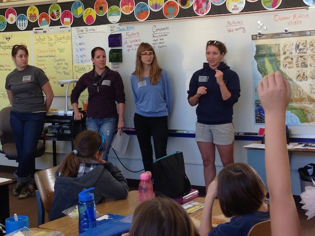
I volunteered for the trip again this year because it is the sort of educational outreach experience that to me really embodies the spirit of MLML; sharing resources and experiences from multiple labs and teaching in our beautiful marine backyard.  The student volunteers represented the Physical Oceanography Lab, the Phycology Lab (Sara Worden), the Benthic Ecology Lab (Dorota Szuta), and the Ichthyology Lab (Heather Kramp). Another reason I volunteered again? Try passing up an opportunity to geek out science on one of the prettiest beaches in the world.  Yeah, it’s tough to do.

Benthic Ecology Lab student Dorota Szuta teaches a group of fourth grade girls about intertidal invertebrates. Photo: Diane Wyse (2013)

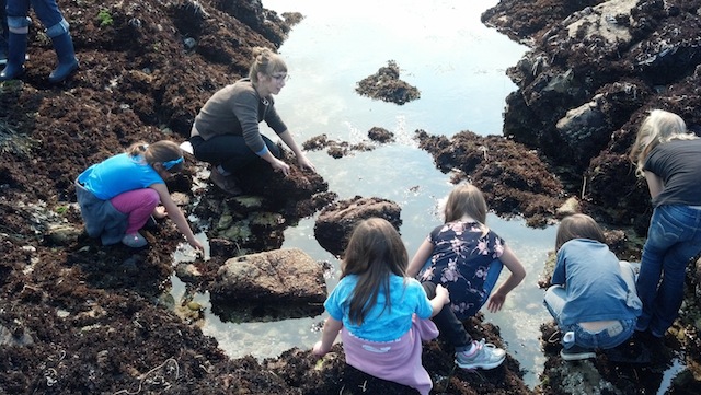
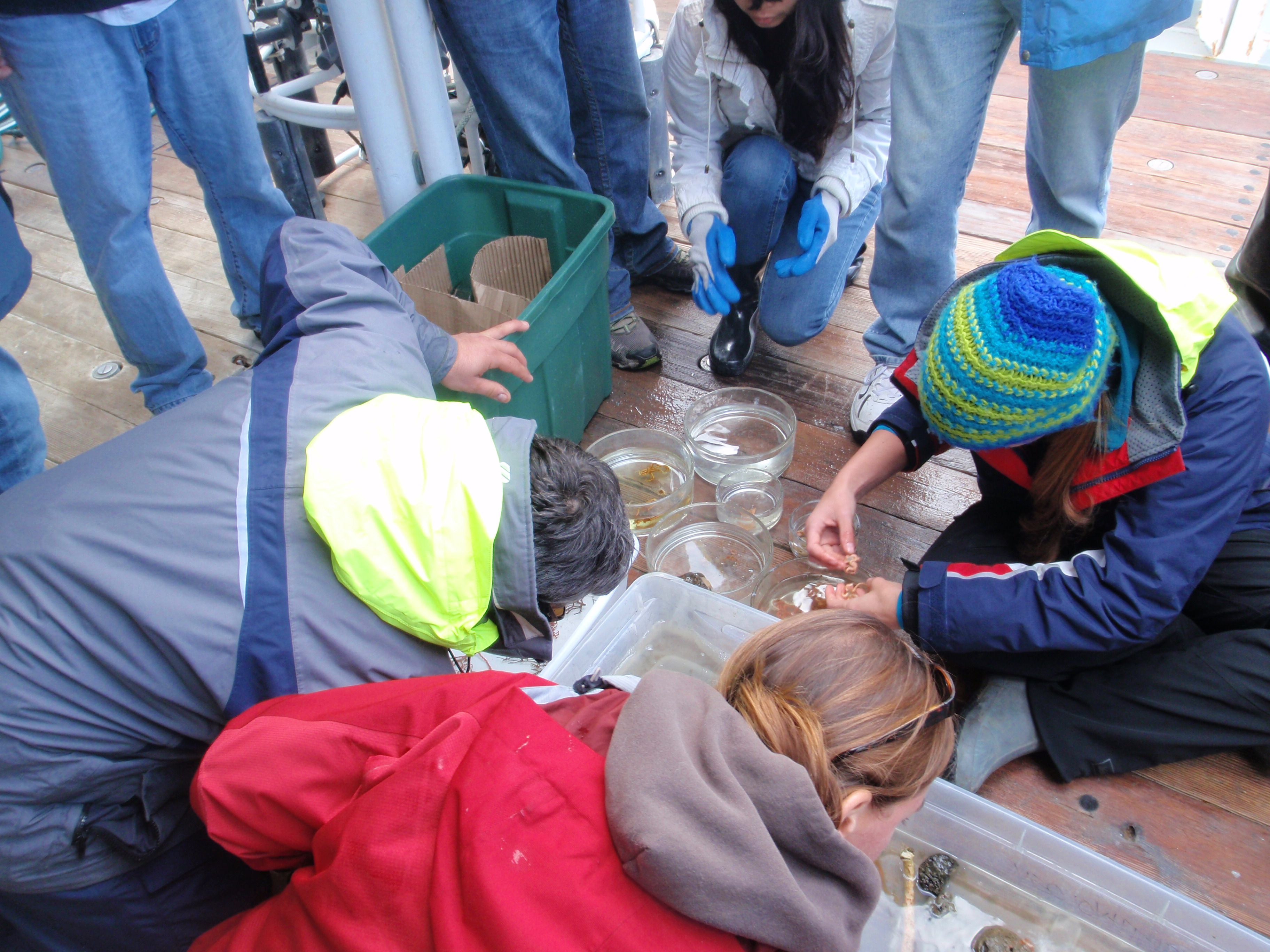
Working off an intertidal food web lesson plan developed by the Teaching Enhancement Program at MLML, the grad student volunteers introduced the fourth grade class to the organisms in tidepools at Asilomar State Beach.  We were impressed by the knowledge the students shared with us that their teacher Alicia Doolittle had introduced in previous lessons.  At the beach it was hard to tell who was more excited to explore the intertidal – the elementary students, grad students, or even the parent chaperones!

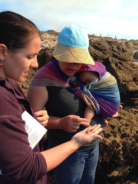
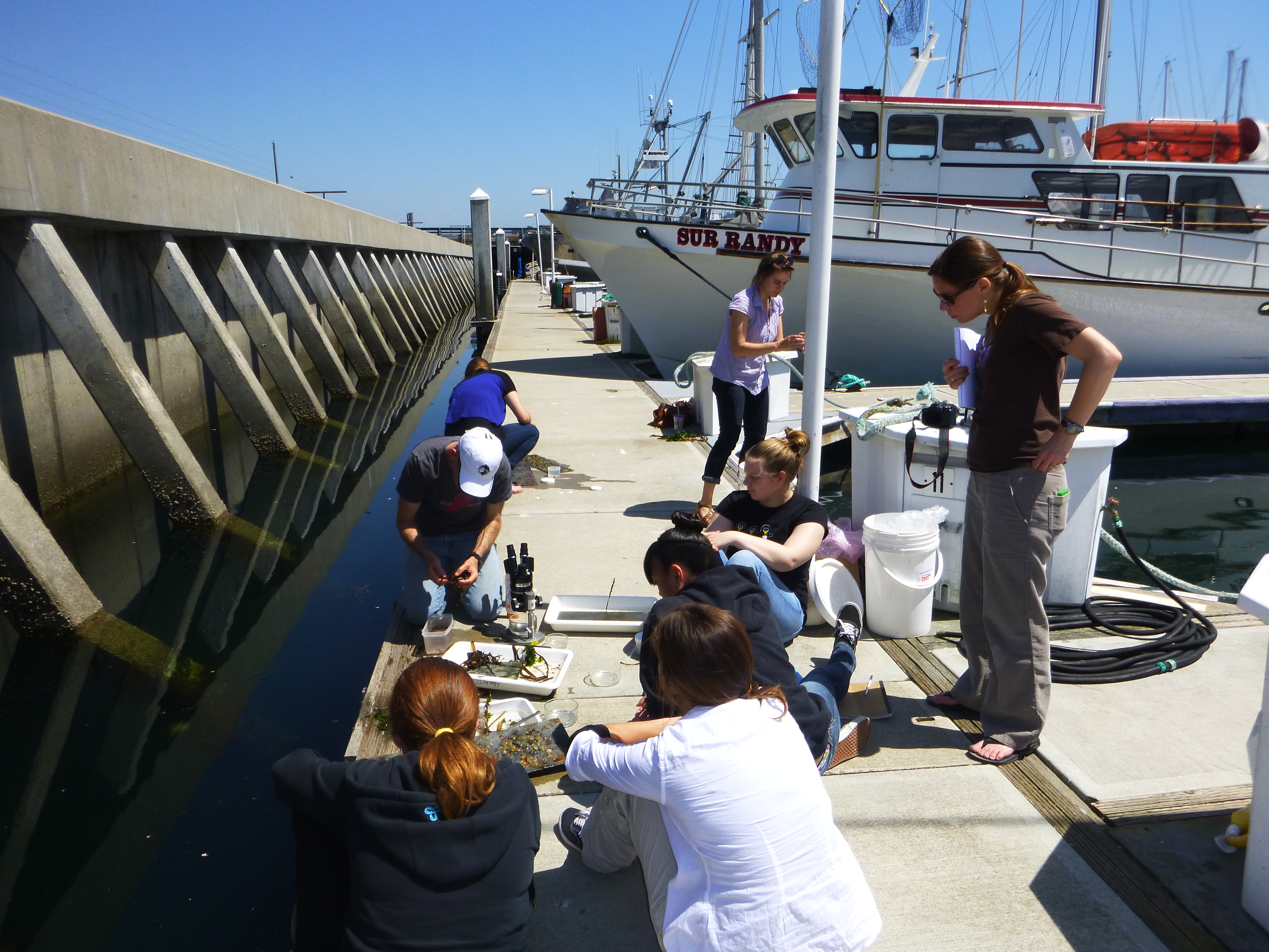
Ichthyology Lab student Heather Kramp shows some intertidal organisms to an interested chaperone and the youngest field trip participant. Photo: Diane Wyse (2013)

This year’s trip was especially cool for me as my graduate advisor, Dr Erika McPhee-Shaw, who serves as a board member for Bay View Academy, was along for the trip and helped to photo-document the field lessons.  I’ll admit it was a bit intimidating to be on the other side of things – here’s a highly-regarded physical oceanographer who has taught me equations of motion, coastal dynamics, and guided me through the steps of a Master’s research thesis, and here I am fielding questions about the inner workings of the ocean to a class of fourth graders while she listens in the audience.  It reinforced something I’ve learned time and again through graduate school, that the more simply and elegantly you can describe a complicated process, the more completely you understand it.  With the students’ healthy appetite for knowledge our conversation ventured from why ocean water is blue to a comparison of ecological zonation on a beach versus a mountain.

Recognize those t-shirts? The 2013 Open House tshirts were designed by our very own Dorota Szuta! Photo: Alicia Doolittle (2013)
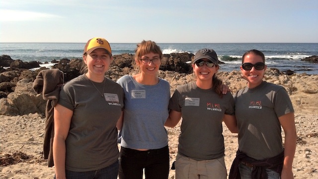
Recognize those t-shirts? The 2013 Open House t-shirts were designed by our very own Dorota Szuta! Photo: Alicia Doolittle (2013)

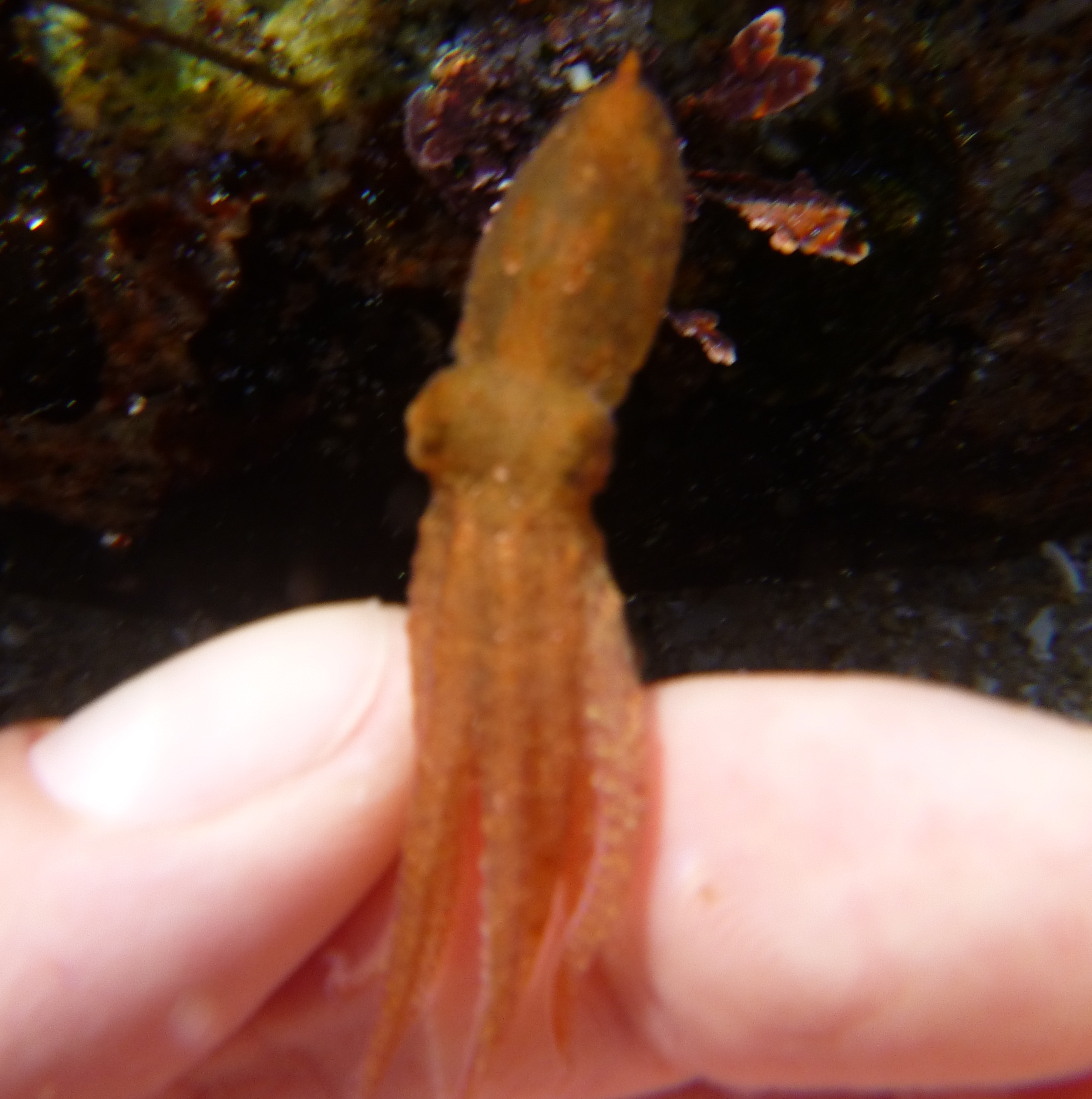
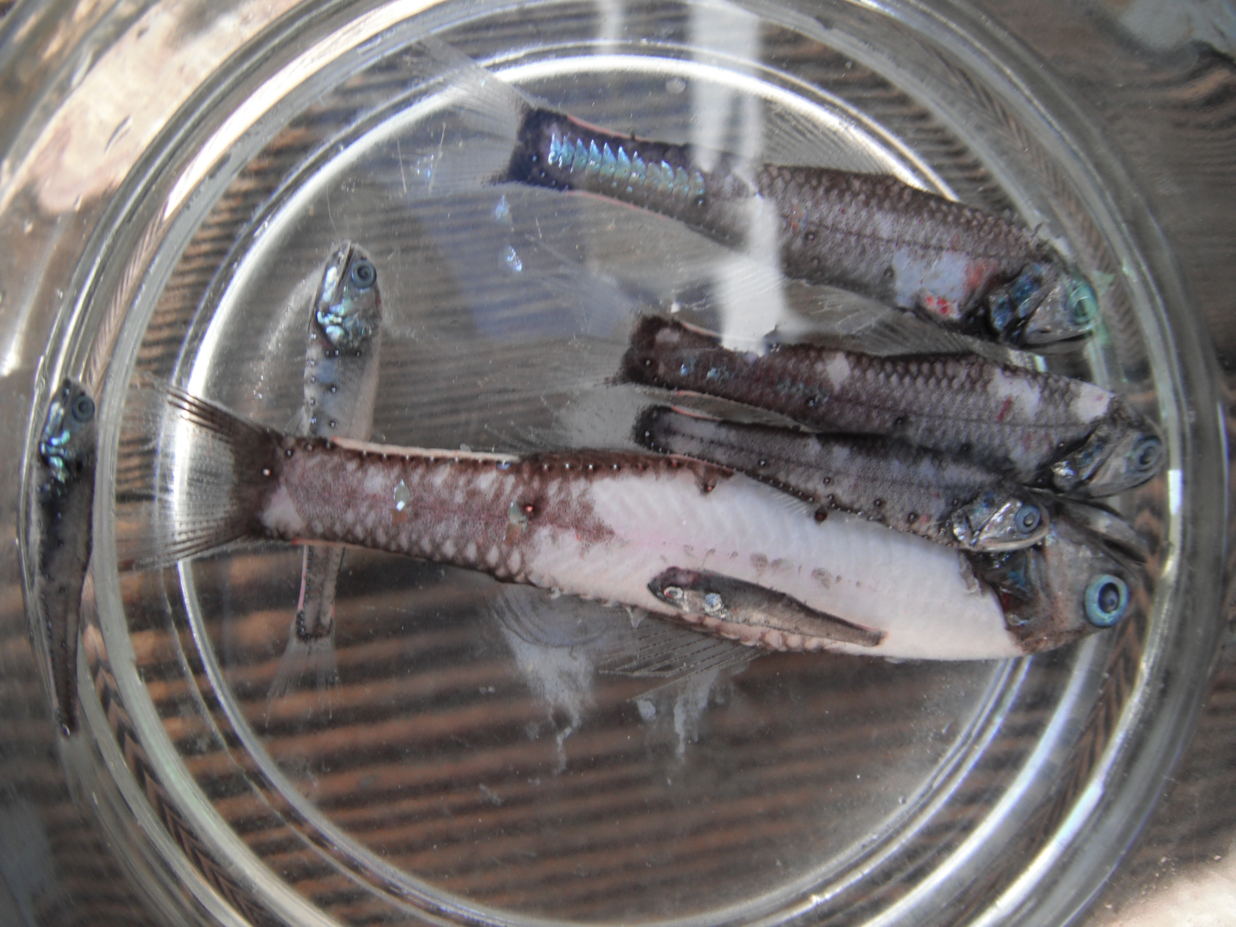
From the closing discussion it was clear that invertebrates were the crowd favorite: the hermit crabs, the purple pisaster (ochre) seastar, even the tunicates were getting some love thanks to the students’ curiosity about the round little chordates.  Will student leaders from MLML lead the trip again?  You better believe it!

Down on the Boardwalk

By Michelle Marraffini, Invertebrate Zoology Lab

Classes at Moss Landing are a lot of work but they can also be a lot of fun.  We get to go on cruises through the bay and learn coastal sampling methods.  This past spring semester the Invertebrate Zoology class, while attempting to learn >30 phyla of invertebrates we took time out to visit my favorite Monterey Harbor.  Here we looked at some PVC settlement plates I had put out earlier in the semester.  They collect may groups of settling animals such as Bryozoans, Chordates (Tunicates), and Barnacles.  Students took time to examine these sessile (not moving or semi-permanently attached) animals under some dissecting microscopes we brought with us.  These sessile animals are part of the marine fouling community, generally known for their ability to attach to almost anything in the water including piers, floating docks (like the one we were standing on in the above picture), and even the bottom of people's boats.  Thanks to a little sunshine and fun things to see, we had a great afternoon on the docks.

Invertebrate Zoology Class
Students sampling settlement plates from Monterey Harbor with Professor Jon Geller (white hat).
Fouling organisms common to floating docks of debris.
Fouling organisms common to floating docks of debris. Note: not all of these animals can be found on the Pacific coast, but below you can see a picture from Monterey Harbor of a few. Illustration by: New England Aquarium


Invasive Tunicates
Local fouling community with many invasive tunicates, clear vase like solitary tunicate (Ciona savangyi); star or flower colonial tunicate (Botryllus schlosseri); and the yellow colonial tunicate in the center and orange tunicate in the bottom left (Botryllodies violaceus).

Invertebrate Spotlight: Larvaceans

By Michelle Marraffini, Invertebrate Zoology Lab

Today in the Marine Invertebrate Zoology we learned about one of the most interesting marine animals.  Larvaceans (Class Larvacean) are unique animals in the phylum Chordata along with their close relatives sea-squirts (Class Ascidiacea) and slightly more distant relatives humans (Subphylum Vertebrata).   These chordates retain their tadpool larva form and excrete a mucus house from specialized cells located on their head.  This house starts off as a small balloon like structure, the tadpole Larvacean whips its body to inflate the balloon with water, then when it is big enough the animal crawls inside, and whips its tail to continue to inflate the house.  Larvaceans will also eat with the help of their house which also contains screens set up to filter water, water is then further filtered by the animal so that it can eat bacteria sized particles.

A schematic of a larvacean in its house with the screens and showing water current flow. Photo Credit: Earthlife.net

They live in this house until the screens become clogged and then they swim out of it start to make a new one.  They discard their old house with sinks to the ocean floor as marine snow.  Marine snow is considered a big source of nutrients to the deep sea, to learn more about how larvaceans contribute to marine snow check out MBARI's website.

http://oceanexplorer.noaa.gov/explorations/02arctic/background/fauna/media/oik_lab.html
Larvacean photo, screens shown in red tint, white folds are more filtering tools, and the animal itself is in the center of it's house. Photo Credit: Arctic Exploration 2002, Per Flood, NOAA/OER

Tis the season for MLML Open House

By Michelle Marraffini, Invertebrate Zoology Lab

The vertebrate ecology lab’s recreation of the inside of a whale. (photo by The Moss Lander).
The Vertebrate Ecology Lab’s recreation of the inside of a whale. (photo by The Moss Lander).

The spring semester is buzzing with activity from classes, field trips, and preparing for Open House.

Have you ever walked inside the belly of a whale?  Want to know how long turtles live or what seastars eat?  This year's Open House will answer these and so many more of your ocean questions.  Be there Saturday April 20th and Sunday April 21st from 9am to 5pm.  As a FREE EVENT we offer a marine adventure puppet show, education presentations by students and faculty, live touch tanks, a sea lion show, raffle and prizes, and so much more.  There is so much to see you will need to come back both days!

Open House!
Entry Way to MLML. Dive into Open House! April 20th and 21st
Photo by Scott Gabara

What’s that on the rock?

By Michelle Marraffini, Invertebrate Zoology Lab

Can you see the octopus?
What do you see on the rock?

What's that on the rock?

The invertebrate zoology class took a field trip to Asilomar State Beach last week to look for cool creatures.  Professor Jon Geller encouraged us to turn over rocks looking for flatworms, the topic of this week's lecture.  As I overturned one rock I noticed something quickly hunker down.  It was this tiny octopus that tried to camouflage itself with the rock.   An octopus's boneless body is well suited for changing its shape and its ability to mimic other animals, algae, and rocks or sand can be quite impressive.  Check out this video of an octopus camouflaging itself ('Where's the Octopus?').  These extraordinary animals are different from other camouflaging animals because they not only change their color and shadow but they also change the texture of their skin to match their background and they do all of this by sight!

Their very kein eyes detect the object they wish to look like and control over 30 million chromatograms (color producing cells) and papilla (cause the three dimensional shape of the skin).  Octopus's do this while color blind which mystifies scientists.

Look under rocks
Small octopus found at Asilomar State Beach hiding under a rock at low tide.

This octopus I found is likely a Pacific red octopus (Octopus reubescens), though it swam away before I could get a good look (no animals were harmed in the making of this blog post).  This is so far the coolest creature I have seen in the intertidal.  Get outside and see what you can find!

Taking off safe and sound
My little buddy swimming away.

What Does a Vampire Squid Really Eat?

By Melinda Wheelock

Researchers at MBARI have discovered that the vampire squid (Vampyroteuthis infernalis) doesn't share a diet with its bloodthirsty namesake. In a recent news release, it was announced that this scary-sounding cephalopod actually feeds on the remains and waste of animals and microalgae that live in shallower waters. This "marine snow" falls through the water column to deeper water, where the vampire squid can pick it up using its unique features. The recent study observed that this creepy creature extends one of its long, thin filaments (which can be 8 times its body length!) to capture floating debris and bring it back towards its body. The vampire squid then scrapes the filament clean with its tentacles, which produce a mucus that sticks the food particles together. This is one 'vampire' that's less scary than it looks!

The vampire squid isn't as bloodthirsty as its name implies.
Photo copyright 1999 Brad SeibelFor more pictures, check out MBARI's web page dedicated to the vampire squid, here.

A Point Sur Adventure

Marine Ecology students on the Point Sur cruise sort and record organisms from the Monterey Bay.

By Kristin Walovich

The Marine Ecology class embarked on a seafaring adventure last Monday on the Moss Landing research vessel the Point Sur to observe the biota of the Monterey Bay. The class was joined by members from the Monterey Bay Aquarium, MBARI and even Professor Emeritus Greg Cailliet who arrived bright and early for a 7am departure time.

After braving choppy water and a bit of rain we began our day with a beam trawl, designed to sample creatures from the ocean floor at 600 meters depth. Unfortunately we were left empty handed when the net returned to the surface with a hole caused from large rocks lodged in the net.

Despite our first strikeout, our second mid-water trawl yielded a wide array of fish, crustaceans, jellyfish, and a plethora of other gelatinous creatures. Once on board the Point Sur, each animal was classified into separate glass dishes and recorded, giving the students a chance to practice their species identification and exercise their Latin nomenclature.

The highlight of the trawl (quite literally) was a group of fish called the Myctophids, or Lanternfish. These fish have light emitting cells called photophores that help camouflage them in the deep ocean waters in which they live. Lanternfish regulate the photophores on their flanks and underside to match the ambient light levels from the surface, rendering them nearly invisible from predators below.

Image
Lanternfish emit light from cells called photophores that help camouflage them from predators.

The last tow of the day was called an otter trawl; but don’t worry, we didn’t catch any sea otters.  This net is name for the ‘otter’ boards positioned at the mouth of the net designed to keep it open as it travels thought the water. The animals are funneled to the back or ‘cod’ end of the net and are brought to the surface for the class to observe.  We saw several species of flatfish including the Sand Dab, Dover and English Sole, several dozen octopuses (or octopodes depending on your dictionary) and even a pacific electric ray.

After a long day of sunshine, high seas and amazing sea creatures the Marine Ecology students were excited with their discoveries, but also ready to be back on solid ground.

Image

Are you my clone?


By Jessica Jang

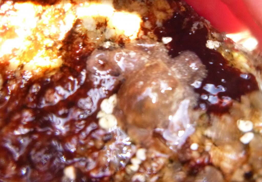
Ever wonder why you see some anemones in groups and some alone in tide pools? Sea anemones can reproduce in two different ways, asexually and sexually. Anemones are broadcast-spawners meaning that they release eggs and sperm into the water column for fertilization.  However if you're an anemone that has settled onto a nice barren rock and don't have time to reproduce, but you want to prevent other anemones  from taking over that rock  you claimed, what do you? You split yourself through........ FISSION! This is asexual reproduction, where the anemone splits itself and creates another one of itself of the exact same genetic material.

Depending on species this process may take days to weeks, but once there are more clones present, more can divide themselves through fission. Sooner or later you'll see whole colonies of anemones on rocks!

Holy moly anemones! Each one of these are identical clones.
Photo courtesy of Catarina Pien

In the intertidal zone one of the limited resources is space for sessile organisms so anemones have adapted a way to populate an area quickly . But what if that pesky neighbor anemone is also asexually reproducing right next to your clones? What would you do? That's when you take drastic measures, by fending them off with your acrorhagi, specialized stinging cells used to deter other anemones from taking over your area.

https://www.youtube.com/watch?v=_jNwWQtLeY4

These battles are intense, both parties may suffer serious damage. As you can see in the video, the anemones when attacked retreat. This is because each one of those tentacles have stinging cells called nematocysts. Animals in the phylum Cnidaria (anemones, corals, jellyfish, and hydrae are part of this group) have these specialized cells.

There is a mechanism that triggers the release of this harpoon-like contraption, when released the harpoon penetrates into the target organism and releases the toxin which is useful to immobilize prey such as fish. If you've ever been stung by a jellyfish that's what exactly is happening; some species of jellyfish such as the box jelly and sea wasp have stings that cause excruciating pain, anemones also have these nematocysts too. However, because our skin is too thick for the nematocyst to penetrate into, you only feel a sticky sensation from touching anemones in the tide pools. The fact that we're immune to most anemone stings in the tide pools doesn't make it acceptable to touch them constantly though, the nematocysts do take quite a lot of energy for these anemones to regulate these mechanism. So the next time you're visiting the tide pools do the anemones a favor and just observe and be amazed at their adaptations for surviving in the intertidal zone!

We maybe small, but please respect us!
Photo courtesy of Catarina Pien

Sponge Behavior & the Emergence of Neural Systems

By Amanda Kahn

In a previous post, entitled "Do sponges have the nerve to eat?", Mr. Singer Singh asked the following question:

"It is found that sponges tend to show different behaviors when exposed to certain stimuli such as touch, air and poison it result in closure of osculum and pores. but then how those response is possible with out any brain or nerves?"

I didn't have all of the background to answer his question, so I forwarded it to Nathan Farrar, a graduate student at the University of Alberta who studies just such behaviors in sponges.  Check out his post below:

Sponge Behavior and the Emergence of Neural Systems

by Nathan Farrar, University of Alberta

This is a very interesting question, in fact, likely one of the more interesting in sponge physiology. It is of course quite true that despite histological searches for nerve or neural-like tissue in sponges, the absence of such tissue is bona fide.  It is also true that sponges exhibit coordinated behaviors in response to diverse stimuli.  For example, Ephydatia muelleri and Spongilla lacustrus, both demosponges, generate an “inflation-contraction”-type behavior.  While a video is worth a thousand words, imagine looking down on a sponge in such a way that the canal system is visible.  During the inflation period, the canals throughout the animal ‘inflate’ allowing the canal system to be engorged with water.  During the contraction phrase, as the name suggests, the canal system is contracted exerting force on the water in the channels thereby forcing it out of the canal system through the osculum (i.e., the vent from which filtered water passes from the animal).  This coordinated behavior serves to flush the canal system of any accumulating debris or toxins, but as the questioner notes can also be triggered by mechanical force.  (See a video of the inflation-contraction response here, http://jeb.biologists.org/content/210/21/3736/suppl/DC1)

So, in short, the facts of the question are entirely correct, but how is this response is generated,  anticlimactic as it may be, is unknown.  A few ways through which behaviors can be coordinated in an organism are via electrical signaling, chemical signaling and mechanical coupling.  I’ll comment here on the first two:  There is one known example of electrical signaling in the form of an action potential in the syncytial glass sponge (Class Hexactinellida), however, the response involved is the arresting of the feeding current, rather than a whole body response as is the case with the “inflation-contraction” response described above.  With respect to chemical signaling, the amino acid L-glutamate has been shown to trigger the “inflation-contraction” response in Ephydatia muelleri in a dose-dependent manner.  Interestingly, in Ephydatia, GABA acts antagonistically with glutamate to suppress the response.  Now, this is curious because glutamate and GABA are major excitatory and inhibitory neurotransmitters, respectively, in animal nervous systems.  Other molecules classically thought of in terms of neurotransmission have also been described in sponges including, serotonin, acetylcholine, epinephrine, norepinephrine, and nitric oxide.  Furthermore, a set of proteins collectively known as post-synaptic density proteins, named for their clustering in neurons, have also been shown to be present in sponges.  What role(s), if any, these other molecules play in coordinating sponge behaviors is unknown.  Furthermore, how glutamate triggers and “inflation-contraction” response, or how GABA inhibits it is unknown.  One hypothesis is that a calcium wave is initiated by glutamate which spreads across the sponge body serving as a coordinating signal for the behavior.

If we consider these facts for a moment we realize there are some interesting evolutionary implications.  Here are a group of animals with no nerves or muscle, yet able to sense their environment and initiate coordinated body responses.  Yet, they also possess a set of “neural” proteins.  While these observations are compatible with more than one hypothesis, one certainly worth examining is that sponges resemble animals situated at the edge of acquiring what we would recognize as a primitive nervous system.

Further reading:

On coordinated behavior in sponges, see Leys, S.P., Meech, R.W. (2006). Physiology of Coordination in Sponges.  Can J Zool. 84: 288-306.

On sponges and the emergence of neural systems, see Renard, E., Vacelet, J., Gazave, E., Lapebie, P., Borchiellini, C., Ereskovsky, A.V. (2009).  Origin of the neuro-sensory system: new and expected insights from sponges. Int Zool. 4: 294-308.

And, Nickel, M.  (2010).  Evolutionary emergence of synaptic nervous systems: what can we learn from the non-synaptic, nerveless Porifera?  Invert Biol. 129: 1-16.