
By Duncan Campbell, MLML Ichthyology Lab
Total solar eclipses over tropical reefs are a rare and exciting occurrence - There will only be 54 more total eclipses this century, and more than half of those will occur near the poles, in the middle of the ocean, or over large landmasses. In Mexico, there will only be 2 more total eclipses this century. The effects of eclipses on land are well documented, but how do animals underwater respond? We knew that we were going to experience a 90% total eclipse during our trip to Isla El Pardito in Baja California Sur for MS 273: Marine Environmental Studies of the Gulf of California and had planned to be on shore watching it with eclipse glasses. However, once we got there a few of us had a more interesting idea. Why not go for a dive during the eclipse? I was diving constantly for my project, but I really didn’t have the time to slow down and explore during my dives. I was excited to observe how animals behaved during a unique celestial event. Plus, how many people can say they went for a dive during a nearly total solar eclipse?
The Eclipse itself!
The idea of an eclipse dive had been bouncing around for a few days, but it mostly just a “wouldn’t it be fun” idea. The night before the eclipse, while we were planning the next day’s activities, I decided that I absolutely had to do it. It was a challenge to plan - we were going out on a boat that morning, and if we were delayed while returning, I might miss my chance. The eclipse peaked at 11:00 AM, and our boat would be getting back between 10:00 and 11:00. Since you always need buddies while you’re diving, I roped in one of my classmates, Jonah, and our TA, Roxy. We brought extra air tanks on our boat and decided that if we were running late, we would dive off the boat to get in the water on time. Fortunately, we timed our return perfectly and managed to get back, set up our gear on the beach, and walk into the water around 10:30.
We had great conditions starting the dive - the water was calm, clear, and warm. We swam south towards one of the deeper reefs surrounding El Pardito. Unlike most of our dives, where we were covered in transect tape reels, data slates, and other scientific equipment, this time we only had dive lights and underwater cameras. We worked our way deeper into the reef, watching and recording as the reef slowly darkened. At 11:00, the eclipse reached its totality, the point where 90+% of the sun is blocked. The water had become noticeably darker than before, and it felt like we were diving at sunset.
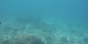
On land, eclipses cause birds to stop singing and send most small animals into hiding. The air gets colder and the light vanishes without a clear cause, an event like no other. However, without a change in temperature or other characteristics, an eclipse is just a change in the light underwater. Plenty of other things, like a storm overhead or dirt in the water, can change the amount of light underwater. It’s hard to tell how much fish respond to the eclipse vs other events as eclipses are a rare occurrence for a comprehensive analysis. To me, it seemed like the fish were more timid than usual, darting away and hiding from us. The schools of tiny wrasses that usually hovered over the corals hid closer to the rocks and inside the heads of corals. The larger fish swam away a little more quickly, and we saw less of the big predator species like snappers that usually patrol the reefs. It may not have been that unusual, but it felt like they noticed that something was different. We took photos and videos, but the dimness and change in environment doesn’t show well on camera.
It was an odd experience seeing how silent the reef felt, even with the constant burble of my scuba gear in the background. It was strangely still and slow, and even after the light started to come back, it was a little subdued. It’s hard to say if the animals really did react to the eclipse in a significant way, but it sure felt like they did. The fish around El Pardito aren’t particularly afraid of scuba divers, and usually swim right up to you. You can practically reach out and touch most fish. Pictures and videos can’t quite do justice to the experience of a sunset dive at noon.
Out of the Eclipse and Into New Environments:
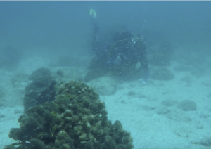
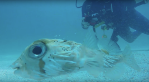
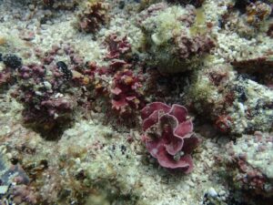
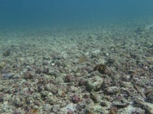
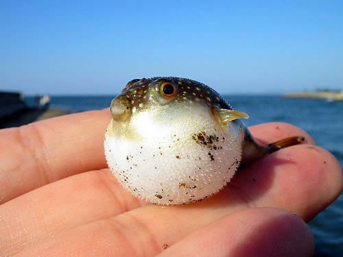
As the eclipse ended, we swam deeper, through the reef towards a large sandy stretch below. We wanted to explore and see what might be down there. To our surprise, we found a rhodolith bed! Rhodoliths are calcareous red algae nodules that form on the bottom of the ocean in dense patches. This bed stretched as far as the eye could see and was full of little pebbly algae and shells. We explored the rhodolith bed for a few minutes before turning around - we had been down for about 25 minutes and only had about 50 minutes of air. As the water brightened back up, we started to see more of the usual suspects on the reef. Brightly colored wrasses schooling above the reef, parrotfish plucking bits of algae off the rocks, leopard groupers cruising around looking for a snack. As we worked our way back across the seafloor towards the island, we started to notice little “piles” of pufferfish. Little groups of pufferfish sleeping on top of each other in full daylight. One of these groups had multiple pufferfish aggregated in an old metal basket underwater, with all the puffers clustered around a scorpionfish. We’re not sure, but we think they went to sleep during the eclipse and hadn’t really woken back up yet. It was a fascinating moment - pufferfish are fairly solitary creatures and we were shocked to see so many in one spot.

Final Thoughts:
Diving during the eclipse was a unique experience and one of the coolest dives I’ve ever been on. It was a great reminder to slow down every once in a while and enjoy the environment you’re working in, instead of just hammering on your work. We found a new rhodolith bed and saw some unusual fish behavior, none of which would have happened if we hadn’t decided to take time to explore and have fun. Our trip to El Pardito was an experience of a lifetime, and the kind of education you can never get in a classroom. I’ll remember my eclipse dive for the rest of my life. It really goes to show the value of a unique experience like MS 273, and how the best education comes from going outside and seeing the world.


.jpg/250px-Spirobranchus_giganteus_(assorted_Christmas_tree_worms).jpg "Christmas Tree Worms")
