8 Days at Sea

By Kelley Andrews, Pacific Shark Research Center

The high-pitched whine of the winch jolts me awake.  I come groggily to my senses, noticing the cigarette smoke from some of the crewmembers wafting through the door of the bunkroom and the dim morning light.  It’s somewhere around 5:45 am.

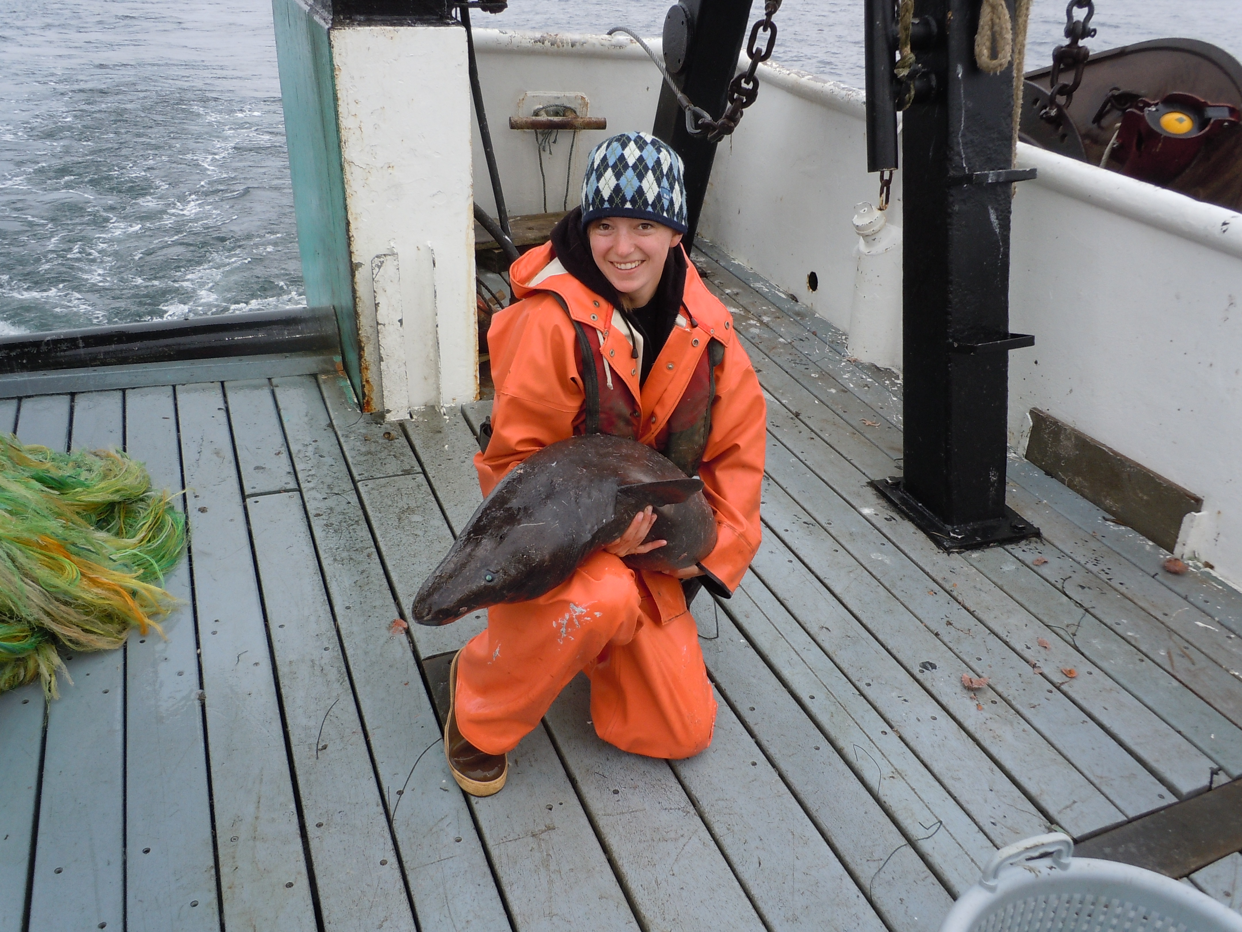
It is my third morning out at sea.  I am on the F/V Noah’s Ark, volunteering for a leg of the National Marine Fisheries Service (NMFS) Fisheries Research Analysis and Monitoring (FRAM) survey.  The mission of the survey is to assess the health of groundfish populations off the west coast of the United States.  The survey makes two passes of the coast from Washington to Southern California every summer, fishing and taking samples and data.  I am part of a team of three scientists, and we are with a crew of four fishermen on the 80-foot vessel.  Right now we are somewhere west of Monterey, CA.

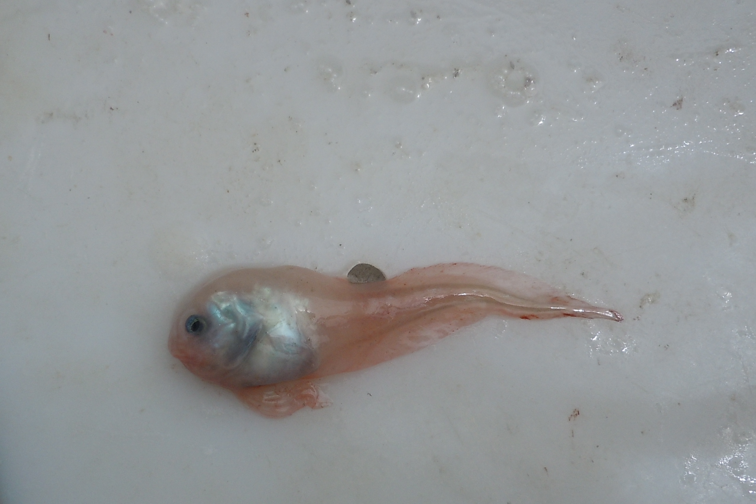
Snail fish.  Photo: K. Andrews (2012)

The first tow of the day begins around 5:30 am, so we can begin processing the catch by 6:30.  The winches deploy and reel in the net from depths over 1,000 feet.  As I go out on deck to get ready to sort fish, I notice that the weather has picked up.  The first two days were flat calm, and I had no idea the ocean could be glassy 50 miles from shore.  But today the winds and swell are picking up, and it feels as though we are headed for rougher weather.

Read More

What Does a Vampire Squid Really Eat?

By Melinda Wheelock

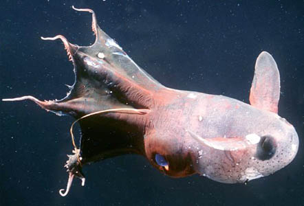
Researchers at MBARI have discovered that the vampire squid (Vampyroteuthis infernalis) doesn't share a diet with its bloodthirsty namesake. In a recent news release, it was announced that this scary-sounding cephalopod actually feeds on the remains and waste of animals and microalgae that live in shallower waters. This "marine snow" falls through the water column to deeper water, where the vampire squid can pick it up using its unique features. The recent study observed that this creepy creature extends one of its long, thin filaments (which can be 8 times its body length!) to capture floating debris and bring it back towards its body. The vampire squid then scrapes the filament clean with its tentacles, which produce a mucus that sticks the food particles together. This is one 'vampire' that's less scary than it looks!

The vampire squid isn't as bloodthirsty as its name implies.
Photo copyright 1999 Brad SeibelFor more pictures, check out MBARI's web page dedicated to the vampire squid, here.

Are you my clone?


By Jessica Jang

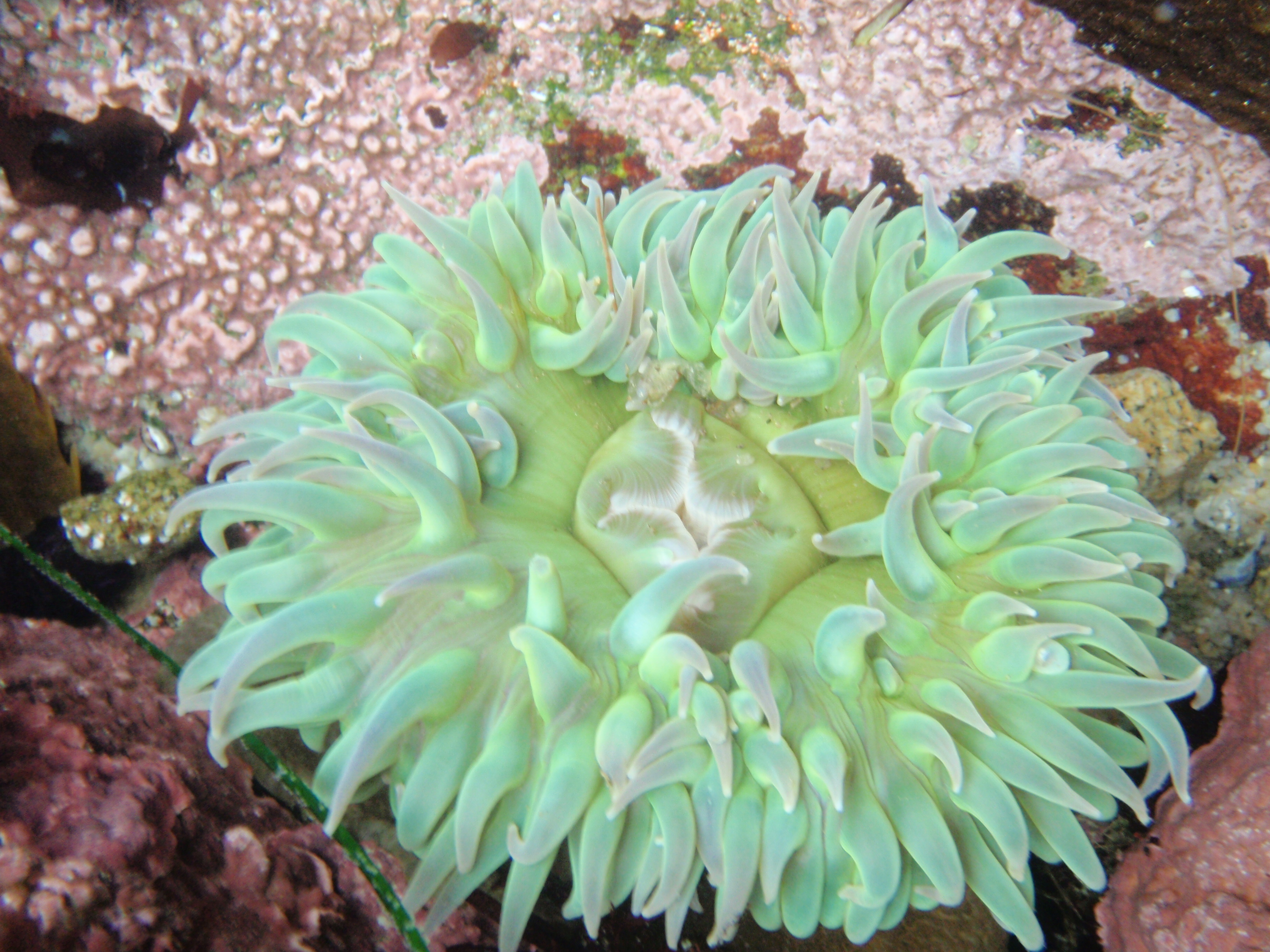
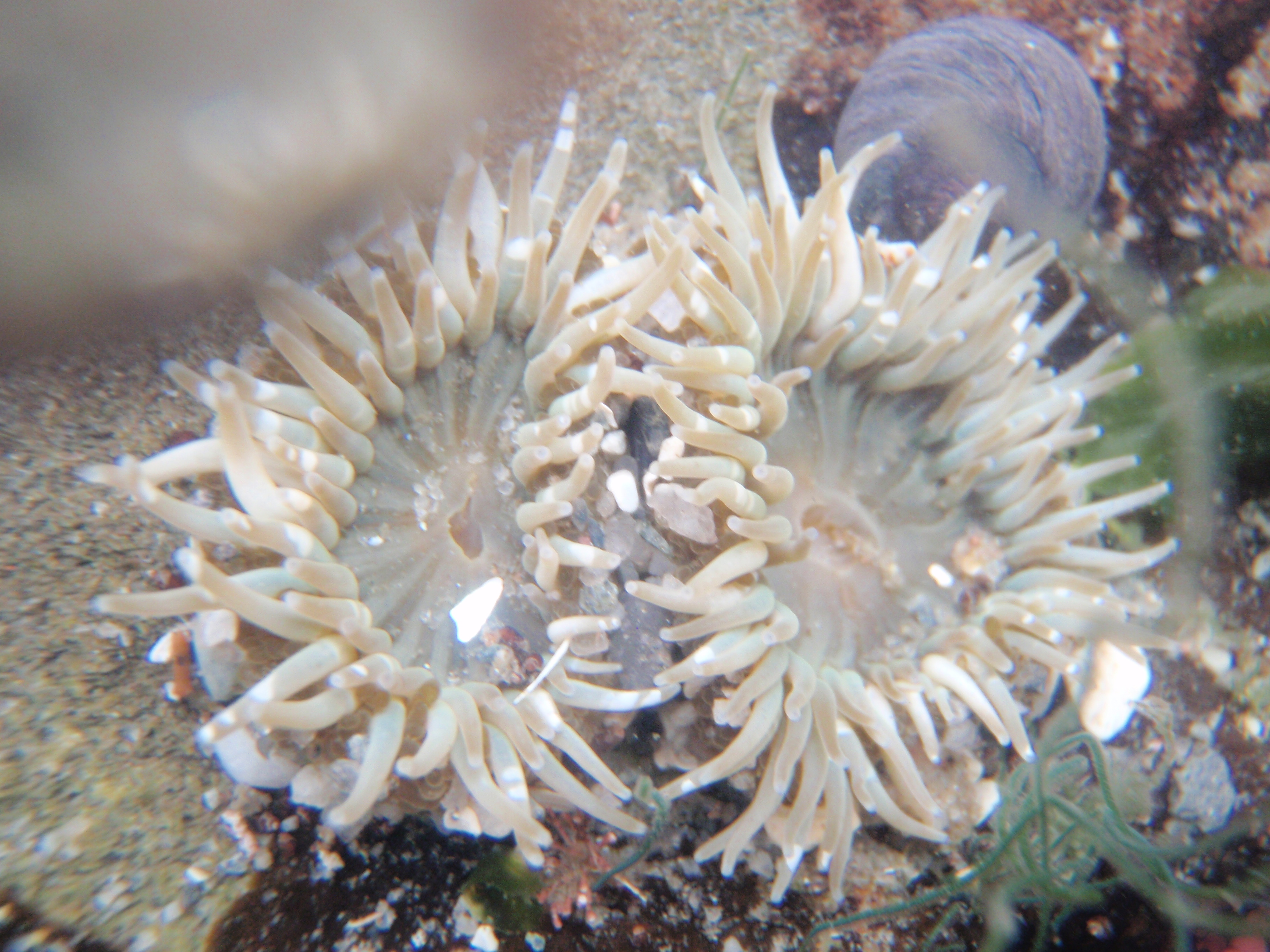
Ever wonder why you see some anemones in groups and some alone in tide pools? Sea anemones can reproduce in two different ways, asexually and sexually. Anemones are broadcast-spawners meaning that they release eggs and sperm into the water column for fertilization.  However if you're an anemone that has settled onto a nice barren rock and don't have time to reproduce, but you want to prevent other anemones  from taking over that rock  you claimed, what do you? You split yourself through........ FISSION! This is asexual reproduction, where the anemone splits itself and creates another one of itself of the exact same genetic material.

Depending on species this process may take days to weeks, but once there are more clones present, more can divide themselves through fission. Sooner or later you'll see whole colonies of anemones on rocks!

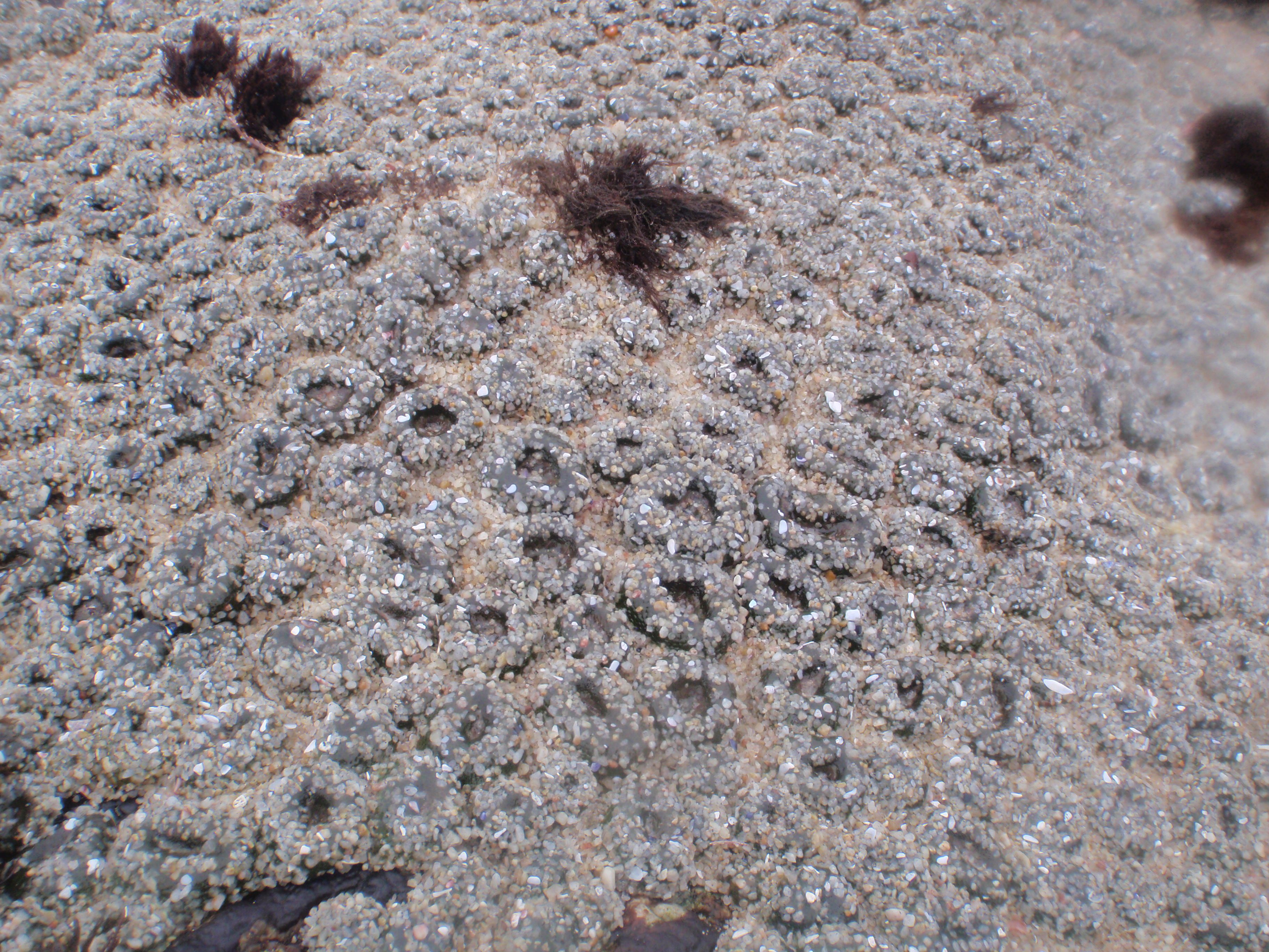
Holy moly anemones! Each one of these are identical clones.
Photo courtesy of Catarina Pien

In the intertidal zone one of the limited resources is space for sessile organisms so anemones have adapted a way to populate an area quickly . But what if that pesky neighbor anemone is also asexually reproducing right next to your clones? What would you do? That's when you take drastic measures, by fending them off with your acrorhagi, specialized stinging cells used to deter other anemones from taking over your area.

https://www.youtube.com/watch?v=_jNwWQtLeY4

These battles are intense, both parties may suffer serious damage. As you can see in the video, the anemones when attacked retreat. This is because each one of those tentacles have stinging cells called nematocysts. Animals in the phylum Cnidaria (anemones, corals, jellyfish, and hydrae are part of this group) have these specialized cells.

There is a mechanism that triggers the release of this harpoon-like contraption, when released the harpoon penetrates into the target organism and releases the toxin which is useful to immobilize prey such as fish. If you've ever been stung by a jellyfish that's what exactly is happening; some species of jellyfish such as the box jelly and sea wasp have stings that cause excruciating pain, anemones also have these nematocysts too. However, because our skin is too thick for the nematocyst to penetrate into, you only feel a sticky sensation from touching anemones in the tide pools. The fact that we're immune to most anemone stings in the tide pools doesn't make it acceptable to touch them constantly though, the nematocysts do take quite a lot of energy for these anemones to regulate these mechanism. So the next time you're visiting the tide pools do the anemones a favor and just observe and be amazed at their adaptations for surviving in the intertidal zone!

We maybe small, but please respect us!
Photo courtesy of Catarina Pien

A Day on the Bay, Biological Oceanography Style

By Heather Fulton-Bennett, Biological Oceanography Lab

The term cruise generally brings to mind tropical weather and luxurious surroundings, but scientific research cruises are much more about long hours of work and only a few brief moments to enjoy the view. As a new student in the Biological Oceanography Laboratory, I was simply excited to get out on the water.

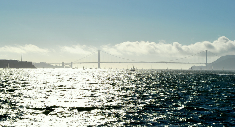
View of San Francisco Bay
Our view of the San Francisco Bay and Golden Gate Bridge as we approached our anchorage for the afternoon

The Biological Oceanography Lab is part of a testing program for ballast water sterilization systems and utilizes the training vessel TS Golden Bear as a semi-mobile research station. With increasing concerns about the spread of invasive species through boating traffic, researchers are trying to minimize the potential for the viable organism to be transported in the ballast water of ships. State regulations focus on minimizing the number of live organisms present following treatment, and our lab is responsible for determining if treatment systems are effective by providing organism counts. Live organism counts are done by microscope on both the untreated and treated ballast water to compare the number of live organisms before and after the treatment. Current regulations require very low numbers of live organisms to be present in the water, so it is crucial to make sure the systems are effective.

Read More

Dem bones, dem dry bones

by Jackie Schwartzstein, Vertebrate Ecology Lab

Most of us remember the song from childhood:

'Toe bone connected to the foot bone, Foot bone connected to the leg bone, Leg bone connected to the knee bone…'

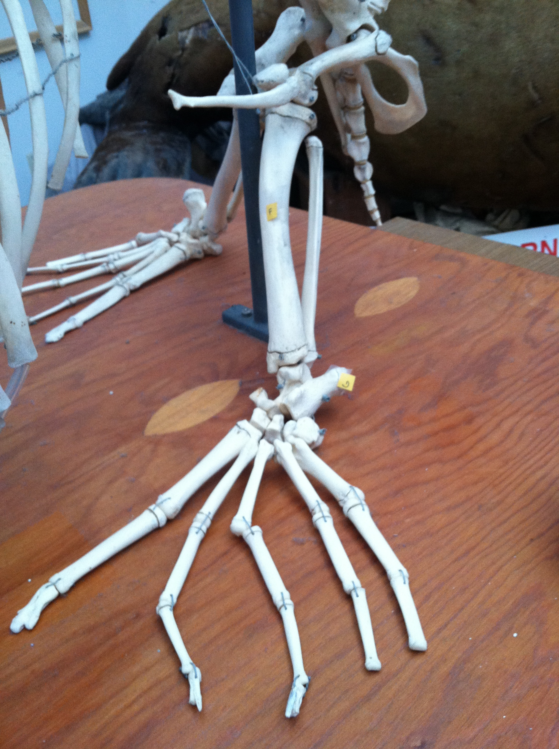
But here at MLML the students in the Marine Birds and Mammals class (MS 112) are quickly finding that what we learned as kids just doesn’t seem to apply anymore! The skeletons of birds, marine mammals, and turtles are MUCH more complicated than the sweet little bones ditty implies. Have the animals changed since I was in fourth grade?! What exactly IS the ‘foot bone’, anyway?!

Rear limbs of the California Sea Lion.
Photo by Jackie Schwartzstein
Can you find the foot bone?

Read More

Pinnacles National Monument

By Gabriela Navas, Invertebrate Zoology Lab

Who would have thought an extinct volcano can be so very multifaceted and interesting? Not me.

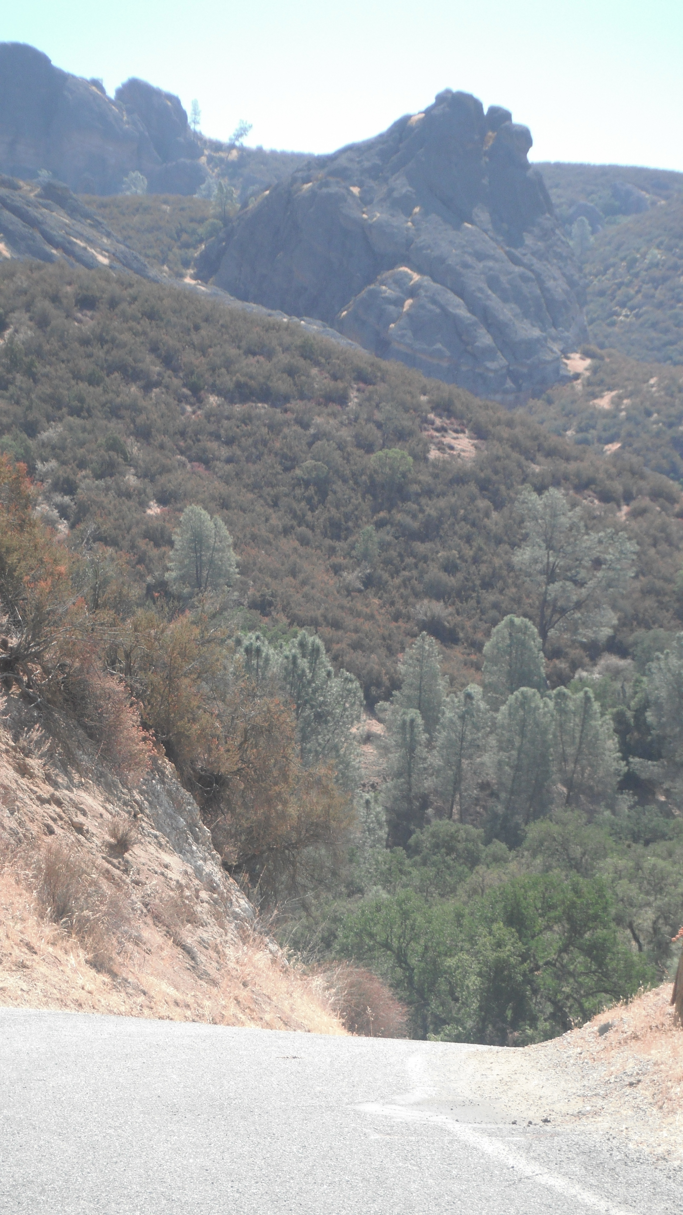
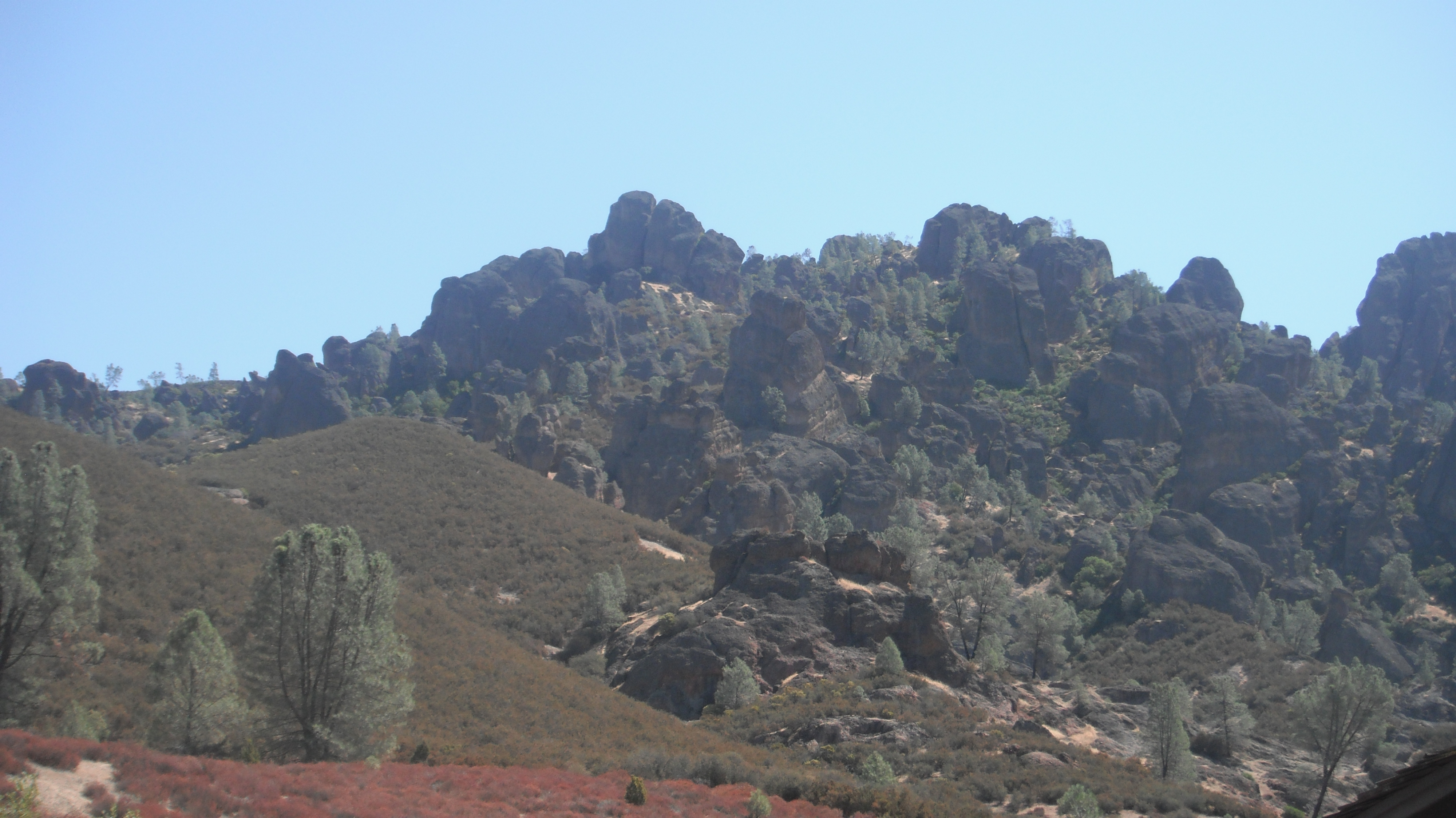
Our Geological Oceanography Class at MLML went to the Pinnacles National Monument the other day, driving up this windy road off of Soledad, California, we see this:

Our professor Ivano Aiello asks the question I very much dread, especially when I have absolutely NO CLUE what the answer could be:

” How do you think these peaks formed that you see in the distance?”

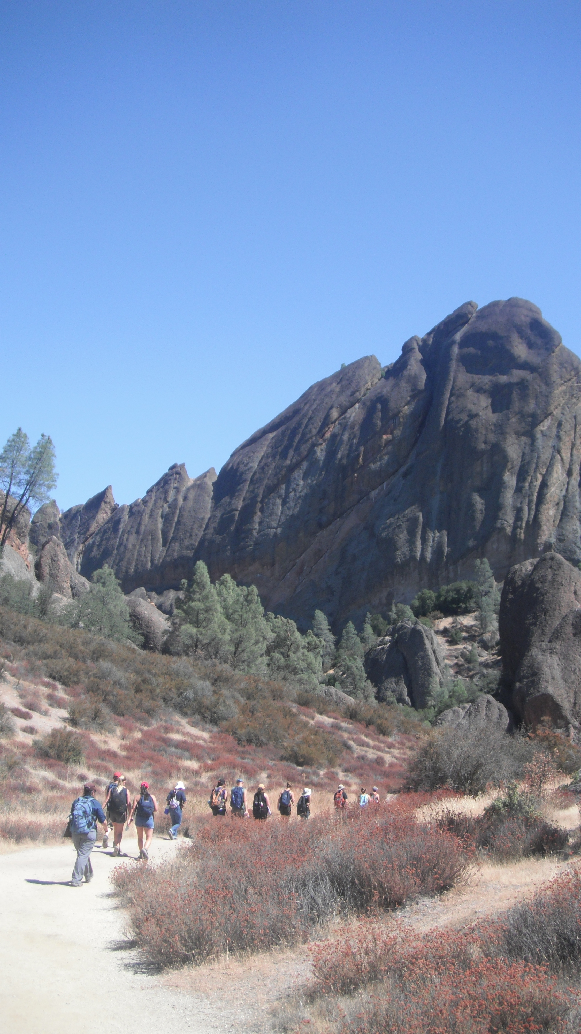
Well, let’s get a bit closer… shall we?

What you see here is actually part of the tube that used to lead into the Magma chamber. The volcano is extinct…no more Magma here, but how did it form?

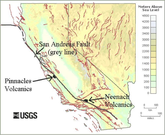
The usual suspects – a fault line, some major earthquake action, you know the Spiel, Bam volcano. Now, the amazing part is that half of the volcano is actually in Southern California as part of the Neenach Volcanics complex, the other up here in Central California. Due to the position of Pinnacles on a fault line it has been transported all the way up north over the period of 30 million years.

Pinnacles features great hiking opportunities, and if you bring a Geologist friend, an amazingly educational hike at that.

Here you see the original side of the volcano. Smoother than the first formations I showed you:

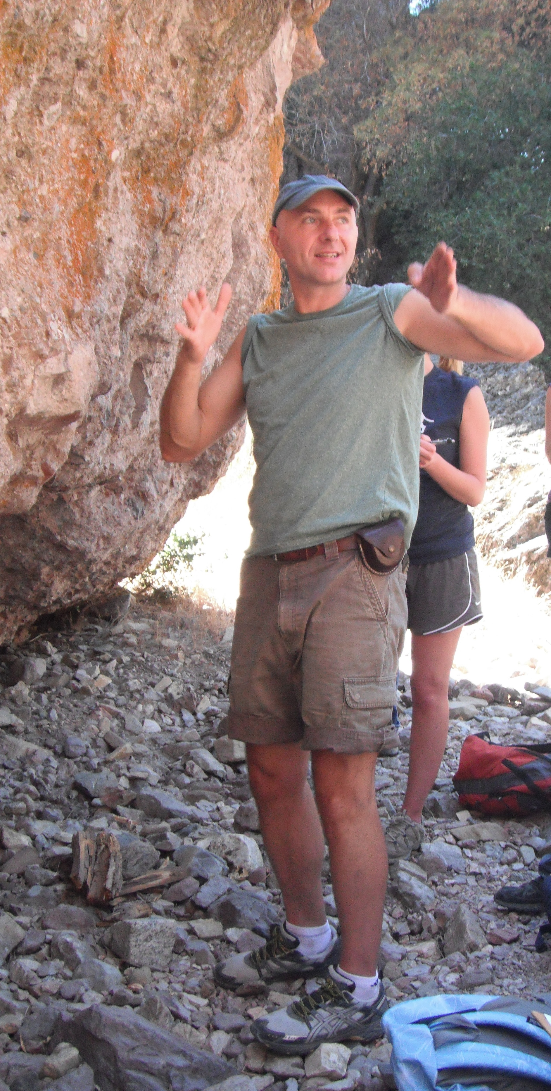
Here’s Ivano explaining one rock formation type at Pinnacles: The Volcanic Breccia, composed of lava flow cementing multiple types of intrusive rocks that originated from the volcano when it was still active.

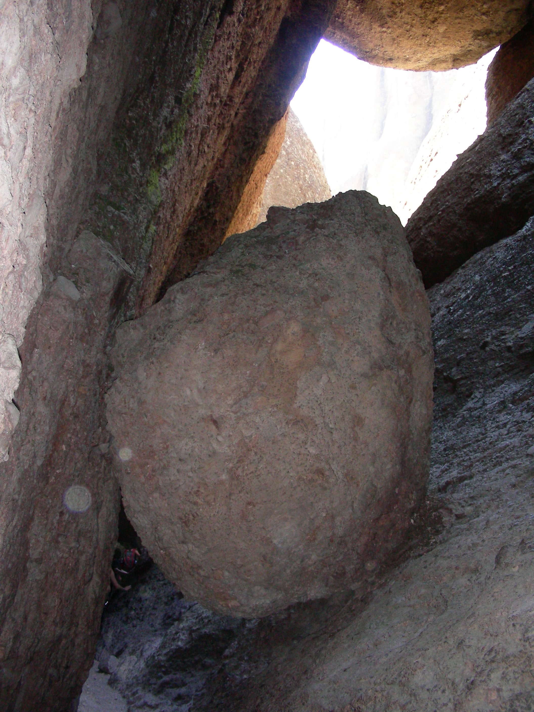
Make sure to bring plenty of water on your hike. The heat can be quite overwhelming. We found huge relief from the heat in caves that were definitely not caves as you may have experienced before. These caves were formed by piled-up boulders that went through some major events from earthquakes to subsequent lava flowing over them, cooling, and some more earthquakes, and an occasional landslide as well, oh sure, floods, too.

Bring your flashlight!

This was a treat!

Sponge Behavior & the Emergence of Neural Systems

By Amanda Kahn

In a previous post, entitled "Do sponges have the nerve to eat?", Mr. Singer Singh asked the following question:

"It is found that sponges tend to show different behaviors when exposed to certain stimuli such as touch, air and poison it result in closure of osculum and pores. but then how those response is possible with out any brain or nerves?"

I didn't have all of the background to answer his question, so I forwarded it to Nathan Farrar, a graduate student at the University of Alberta who studies just such behaviors in sponges.  Check out his post below:

Sponge Behavior and the Emergence of Neural Systems

by Nathan Farrar, University of Alberta

This is a very interesting question, in fact, likely one of the more interesting in sponge physiology. It is of course quite true that despite histological searches for nerve or neural-like tissue in sponges, the absence of such tissue is bona fide.  It is also true that sponges exhibit coordinated behaviors in response to diverse stimuli.  For example, Ephydatia muelleri and Spongilla lacustrus, both demosponges, generate an “inflation-contraction”-type behavior.  While a video is worth a thousand words, imagine looking down on a sponge in such a way that the canal system is visible.  During the inflation period, the canals throughout the animal ‘inflate’ allowing the canal system to be engorged with water.  During the contraction phrase, as the name suggests, the canal system is contracted exerting force on the water in the channels thereby forcing it out of the canal system through the osculum (i.e., the vent from which filtered water passes from the animal).  This coordinated behavior serves to flush the canal system of any accumulating debris or toxins, but as the questioner notes can also be triggered by mechanical force.  (See a video of the inflation-contraction response here, http://jeb.biologists.org/content/210/21/3736/suppl/DC1)

So, in short, the facts of the question are entirely correct, but how is this response is generated,  anticlimactic as it may be, is unknown.  A few ways through which behaviors can be coordinated in an organism are via electrical signaling, chemical signaling and mechanical coupling.  I’ll comment here on the first two:  There is one known example of electrical signaling in the form of an action potential in the syncytial glass sponge (Class Hexactinellida), however, the response involved is the arresting of the feeding current, rather than a whole body response as is the case with the “inflation-contraction” response described above.  With respect to chemical signaling, the amino acid L-glutamate has been shown to trigger the “inflation-contraction” response in Ephydatia muelleri in a dose-dependent manner.  Interestingly, in Ephydatia, GABA acts antagonistically with glutamate to suppress the response.  Now, this is curious because glutamate and GABA are major excitatory and inhibitory neurotransmitters, respectively, in animal nervous systems.  Other molecules classically thought of in terms of neurotransmission have also been described in sponges including, serotonin, acetylcholine, epinephrine, norepinephrine, and nitric oxide.  Furthermore, a set of proteins collectively known as post-synaptic density proteins, named for their clustering in neurons, have also been shown to be present in sponges.  What role(s), if any, these other molecules play in coordinating sponge behaviors is unknown.  Furthermore, how glutamate triggers and “inflation-contraction” response, or how GABA inhibits it is unknown.  One hypothesis is that a calcium wave is initiated by glutamate which spreads across the sponge body serving as a coordinating signal for the behavior.

If we consider these facts for a moment we realize there are some interesting evolutionary implications.  Here are a group of animals with no nerves or muscle, yet able to sense their environment and initiate coordinated body responses.  Yet, they also possess a set of “neural” proteins.  While these observations are compatible with more than one hypothesis, one certainly worth examining is that sponges resemble animals situated at the edge of acquiring what we would recognize as a primitive nervous system.

Further reading:

On coordinated behavior in sponges, see Leys, S.P., Meech, R.W. (2006). Physiology of Coordination in Sponges.  Can J Zool. 84: 288-306.

On sponges and the emergence of neural systems, see Renard, E., Vacelet, J., Gazave, E., Lapebie, P., Borchiellini, C., Ereskovsky, A.V. (2009).  Origin of the neuro-sensory system: new and expected insights from sponges. Int Zool. 4: 294-308.

And, Nickel, M.  (2010).  Evolutionary emergence of synaptic nervous systems: what can we learn from the non-synaptic, nerveless Porifera?  Invert Biol. 129: 1-16.
Diving Adventures in Big Creek

By Catherine Drake, Invertebrate Zoology Lab

For many graduate students at MLML, diving is an essential component to their thesis work, whether it involves collecting samples, obtaining data, or making observations about subtidal ecosystems.  Students must be research dive certified in order to perform these science-related activities.  Here at the lab, we have an excellent research diving program run by our research faculty member and Diving Safety Officer (DSO) Diana Steller. Through this program, students have the option of taking the course either during the fall semester or during a two-week intensive course in the summer.

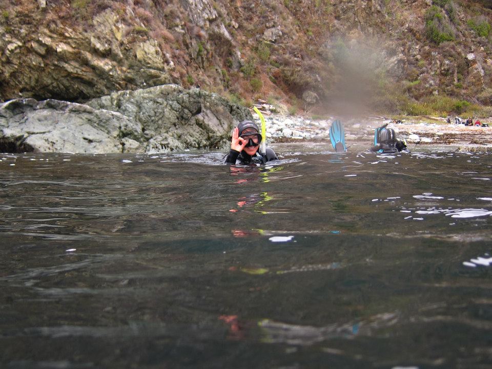
DSO Diana Steller gives the ok after a tough beach entry at Big Creek. Photo by Maria Kyong.

Having gotten my open water certification earlier this spring, I was excited to take the summer research diving class.  For the first week, we practiced a series of underwater skills and swim tests to ensure that we felt comfortable in the water.  There are certain basic scientific skills that must be practiced and perfected to become certified in research diving. These skills include laying out a transect tape and taking observations along the tape.  To master this, we all studied the local fish, invertebrates, and seaweeds to take surveys within the kelp bed for an organization called Reef Check.

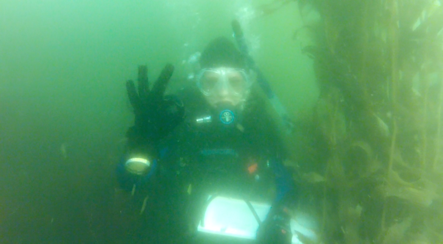
I give the ok signal as I practice a Reef Check survey at Breakwater in Cannery Row. Photo by Scott Gabara.

The following week, we caravanned south to Big Creek State Marine Reserve; while there, we camped in the redwoods and dove consecutively for four days.  We would wake up each morning bright and early, eat breakfast to fuel us for the first dive of the day, and then head to the beach.  Diana and Assistant DSO Scott Gabara would brief us on the dives, we’d suit up and enter the water ready to take data.  After our first dive, we’d sit on the beach with our lunches and warm up in the sun before heading out for our second dive.  Once we completed our second dive, we would wade into the large creek (hence the campsite's namesake), wash off our gear and relax.

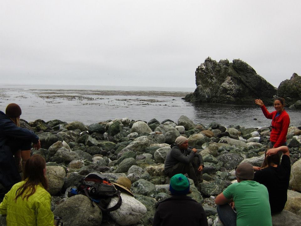
Diana Steller gives a brief on the dive site. Information in this meeting includes beach entry strategies, transect locations, and allowed depths and dive times. Photo by Maria Kyong.

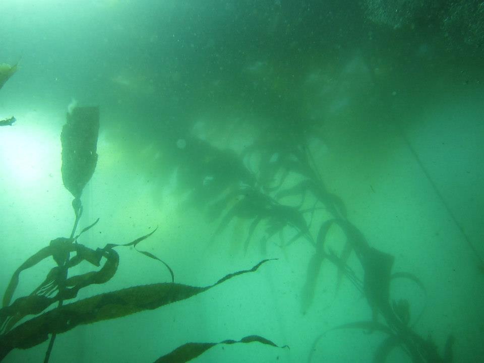
The kelp canopy and sub-canopy are magnificent habitats at Big Creek.  As I swam out to the location of each transect, I’d get entangled in giant kelp (Macrocystis pyrifera) and feather boa kelp (Egregia menziesii), and would use bull kelp (Nereocystis luetkeana) as an anchor when being pushed around by the swell.  Once we descended, the seafloor was inundated with Pterogophora californica and Laminaria setchelii, so much so that I could not see the bedrock below.  To obtain data for Reef Check, we placed the transect under the sub-canopy and crawled our way through the kelp to count stipes, look for inverts, and point our flashlights at unsuspecting rockfish.

Light can barely penetrate the dense canopy of Macrocystis pyrifera and Nereocystis luetkeana. Photo by Marina Kyong.

I noticed that during any dive, something can and will go wrong, especially when you have transect tapes, slates, compasses, dive computers connected to you as you maneuver underwater.  The most important lesson I learned from Diana on this trip was that it’s how you react to these situations that determines your competence and confidence as a research diver.  If you stay calm and remember to always breathe while your mask fills with water, you get caught in kelp, your datasheet falls off your slate, and the surge inverts you, then you are definitely ready for research diving!

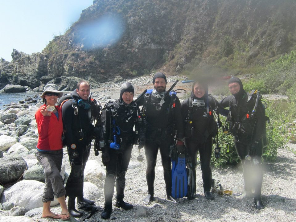
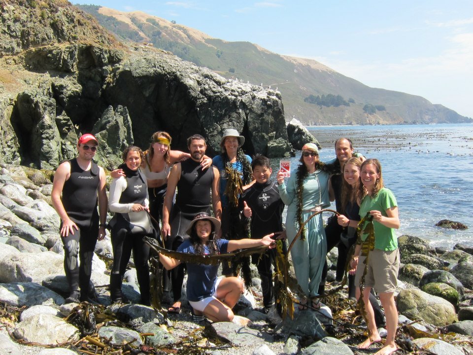
Dive buddies pair up for one last picture after our last, and deepest, dive of the week. Photo by Maria Kyong.
Our awesome summer research diving class! Photo by Maria Kyong.

New Semester, New Students, New Stories

By Diane Wyse, Physical Oceanography Lab

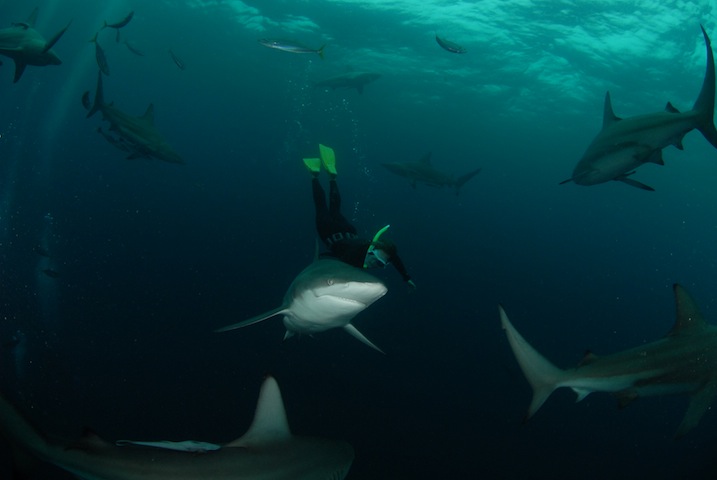
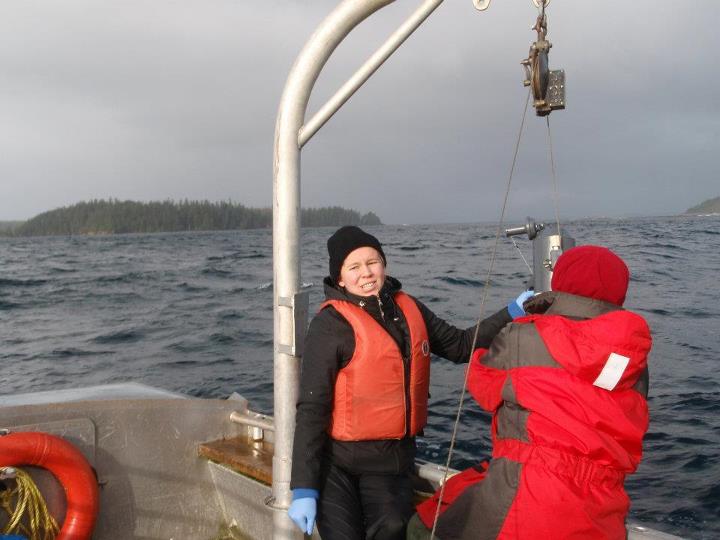
With classes underway, the lab is abuzz with new activities and learning.  This fall, the MLML community welcomes 22 new students to ten of our labs.  Ever find yourself wondering how graduate students at Moss Landing got their start in marine science?  Our new student backgrounds range from gray whale surveys off the Washington coast, to photographing white sharks in South Africa, to shipboard oceanography in Canada, and much more!

Jackie surveying whales off the Washington coast
Kristin freediving in South Africa
Heather performing field research in Canada

Stay tuned for their stories and more from your MLML blog team.

Adventures in Madagascar or On The Importance of Doing a Pilot Study!

by Angela Szesciorka, Vertebrate Ecology Lab

This summer I hopped on a plane, flying 29 hours one way (via Paris — ooh la la) over a period of three days to spend nearly a month on the island of Madagascar working on my pilot study.

Madagascar, a former French colony until 1960, is the fourth largest island in the world. Don’t let it fool you. It looks so tiny next to Africa, but it has 44 percent more area than California, and boasts more than 4,800 km of coastline.

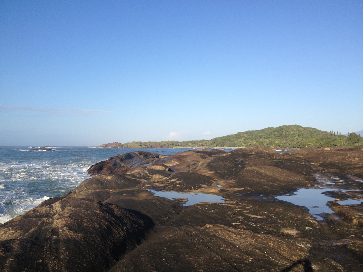
Rocky coastline in Madagascar. Photo by Angela Szesciorka.

Most of the country's export revenue comes from textiles, fish/shellfish, vanilla, and cloves. Newer sources of income include tourism, agriculture, and extracted materials (titanium ore, chromite, coal, iron, cobalt, copper and nickel). Madagascar provides half of the world's supply of sapphires! But with a GDP of around $20 billion, The Economist rated Madagascar as the worst economy in 2011. Most of Madagascar's inhabitants are subsistence livers, meaning they live off of what they can grow or catch.

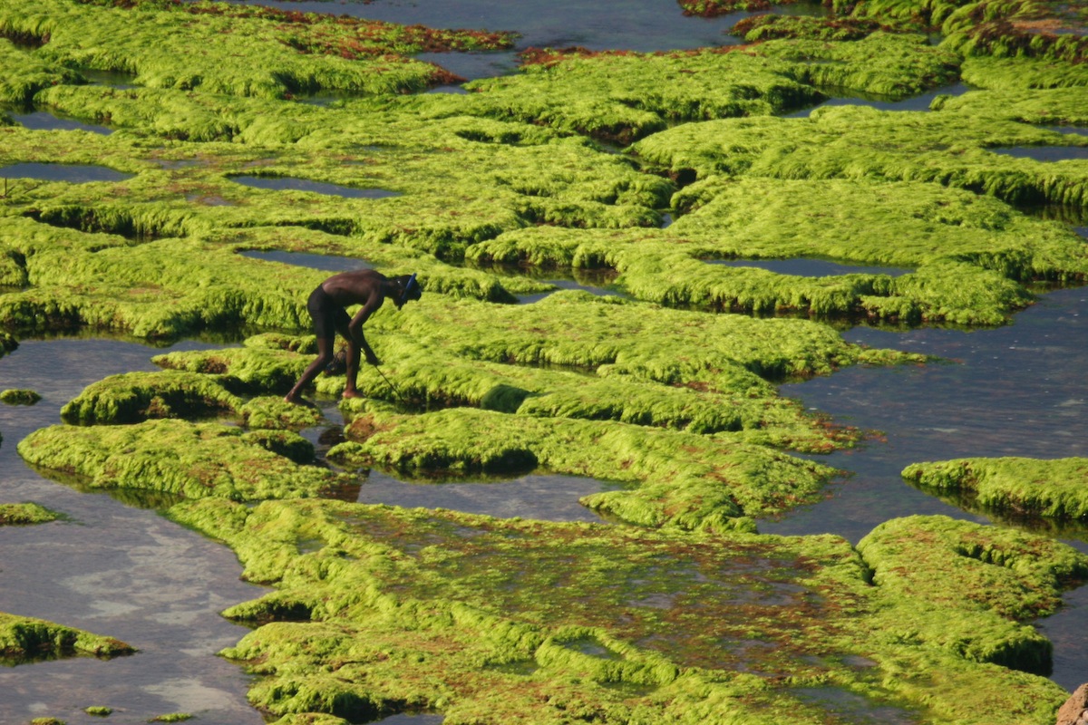
Local fisherman spear hunting for crabs. Photo by Angela Szesciorka.

Read More