The birds and the bees according to coralline crust

Have you ever wondered how algae reproduce? Algal species do not reproduce like plants specifically by using pollen and most do not have special friends like bees to help them reproduce with other individuals. The cool thing about algae is that each kind of algae and sometimes even individual species experience the "birds and the bees" in a different way. For instance, there are some species of algae that belong in the green group that have positive and negative phototaxis gametes. This means that when the gametes (male or female) need to find a mate, they are attracted to the light and come to the surface (positive phototaxis), where they meet all the other singles in the area. Kind of like going out to a bar. Once they have found their mate, they sink to the bottom (due to negative phototaxis) to settle together on a rock and make a new individual. Kelps, belonging to the brown algal group, have special pheromones and flagella that are used in the reproductive process. Female gametophytes or eggs have different kinds of pheromones like animals do that attract the sperm to the egg. Essentially, kelps make the boys do all the work! The males have flagella, or swimming appendages, that help them move towards the female once they recognize the pheromone. Once the female gametophyte is fertilized the female is taken over and the new individual actually grows from that female egg. Red algal species are also very different in how they reproduce.

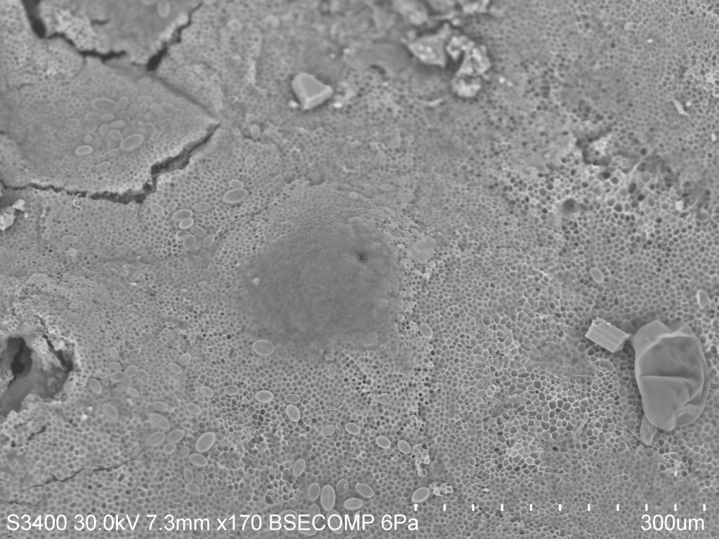
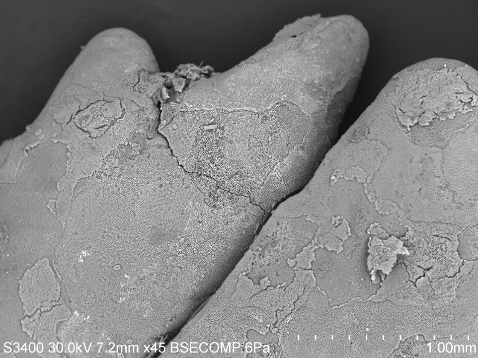
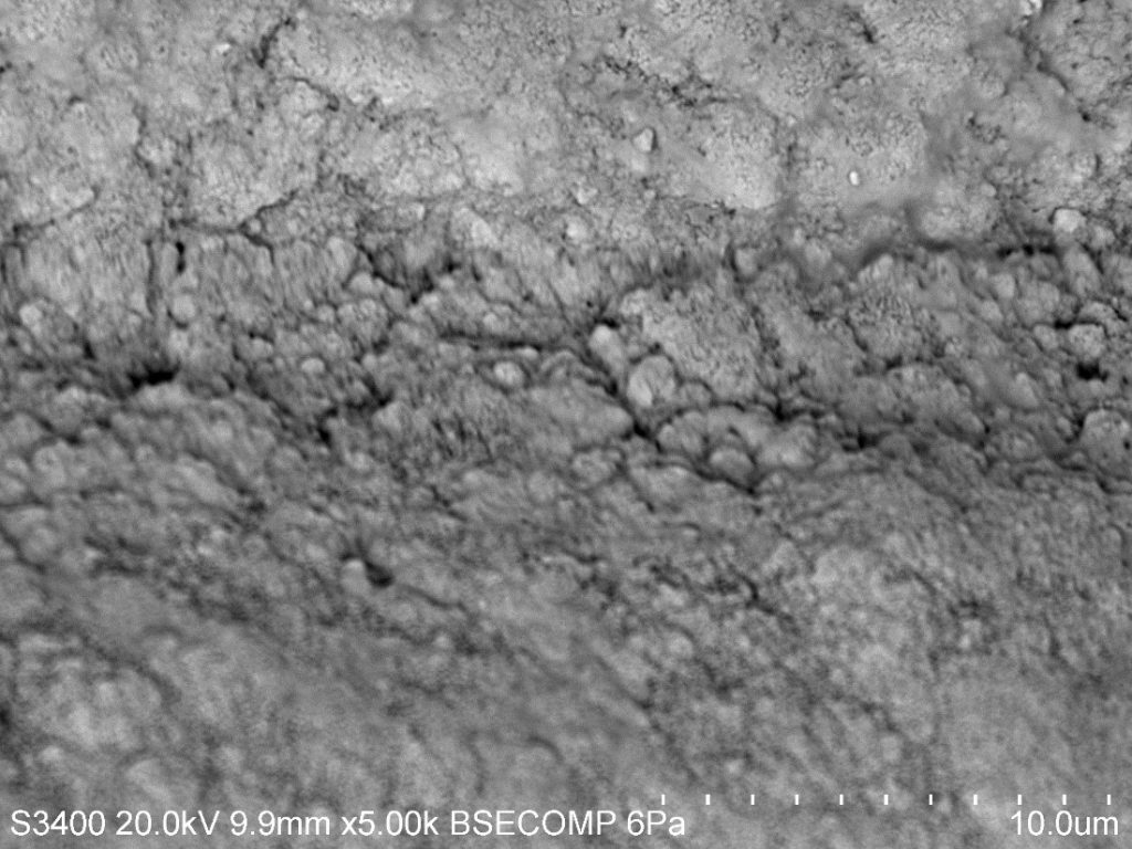
Specifically, coralline algae, the crustose form, has to get very creative in its reproductive cycle. When the coralline crust is ready to reproduce it creates little cenceptacles or cases along the top of the crust. It then fills the conceptacle with reproductive material or tetrasporangia. When they are ready to release this material, the conceptacle bursts through the top layer of the crust and the reproductive material, or tetraspores are cast into the water column to find a mate and settle on a nearby rock before something comes along and eats it! In the picture to the right you can see this conceptacle in the crust. It has yet to fully burst.


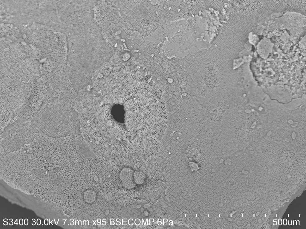
However, the picture on above is on the same piece of crust just in a different area and you can definitely see where the conceptacle used to be. If you were to look at this crust with the naked eye you would not have been able to see any of this. But thanks to the SEM, a human can actually see the detail of an individual conceptacle. To a phycologist, this is SUPER cool! Hopefully you all find it slightly fascinating too!

Piston Core 26

Previous posts in the New Guinea region have discussed sites adjacent to delta fronts. Nearshore sediments are typically variable in nature consisting of sand, mud, pebble-sized particles, and biogenic material. Here, we will focus on piston core 26 (PC26), a drill site furthest from the shoreline.  PC26 was extracted south of Manus Island at a water depth of 875 m and a subsurface depth of 48-49 cm. Sediment deposition in deep water environments, located on the abyssal plain, build up at reduced rates. These decreased rates of sediment accumulation are due to the limiting factors of particle transport; volcanic ash and windborne particles are slowly transported towards the open ocean environment. Therefore, we should expect sediment extracted from PC26 to be largely homogenous.


Immediately, it is apparent that sediment from PC26 is predominantly biogenic material. The image above highlights several chambered tests, or shells of foraminifera. Their test are typically cemented with sand grains or other materials, and crystalline CaCO3 in the form of calcite or aragonite depending on the species. This particular species has large pores and exhibits a trochospiral chamber arrangement. A mature foraminifera may range from 100 µm to 200 cm in length. There are approximately 4,000 species of forams, although only a few modern species are in existence, making these microogranisms the most abundant shelled protists in the marine environment since the Cambrian period.


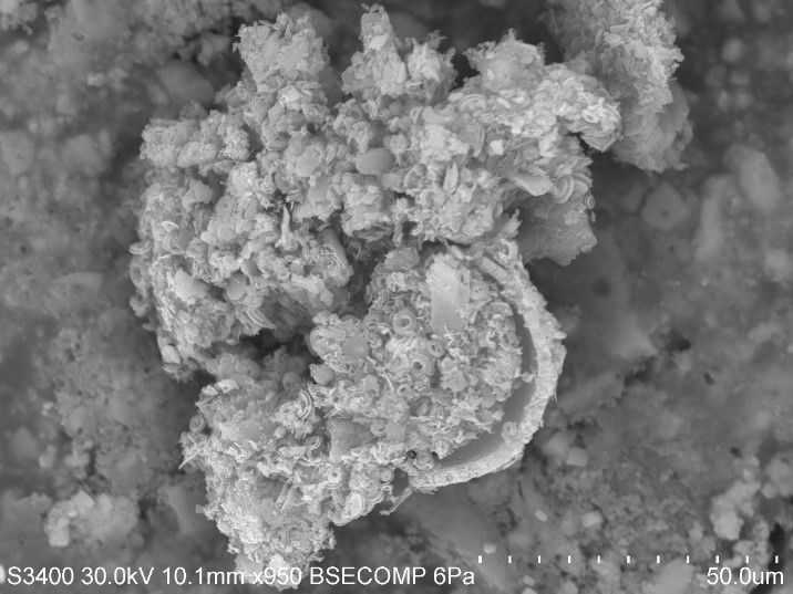
At close inspection, we can see an aggregate of coccolithophores settled within a foraminifera test. There is a distinctive difference in the size and shape of these two microorganisms that have the identical chemical compositions. Therefore, it is suitable to characterize this pelagic sediment as calcareous ooze, given its calcic nature. The remarkably small size of coccolithophores in comparison to forams does not limit its abundance in pelagic sediments. Calcareous oozes cover approximately one-thirds of the Earth’s entire surface, and due to seawater/carbonate interactions, calcareous ooze begins to dissolve below the calcium carbonate lysocline in the water column. Consequently, material below the calcium carbonate compensation depth calcareous ooze completely dissolves.


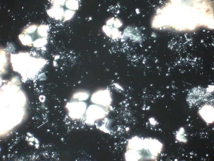
A visual representation of what we see underneath the SEM is also visible in PPL (top) and XPL (bottom). In PPL, the pore-rich tests of the foraminifera are easily distinguishable, particularly in the upper left corner. In XPL their calcite bearing shells illuminate the field of view with their four-chambered anatomy. Similarly, aggregates of coccolithophores are distinguishable in XPL.

Hydroid

Today, we're going to look at a member of the Cniderian phylum, named after the Greek word for "nettle" in reference to their special stinging cells, the nematocyst. You probably think of jelly fish when you think of stinging sea creatures, but Cniderians consist of four subgroups: the Scyphozoa (jellies), the Cubozoa (box jellies), the Anthozoa (sea anemones), and Hydrozoa (hydriods).

Most hydrozoans are fairly inconspicuous. You probably know them as the fuzzy masses you see growing on pilings and boat-bottoms. While most are fairly harmless, there are some capable of delivering a fairly painful sting. For this discussion we're going to be focusing on colonial hydroids. In these communal groupings, individual polyps are connected and share resources through a hydrocaulus. Colonial hydriods have specialized individuals, or "zooids," that exist to fulfill a specific need in the colony. For example, the most common type of zooid is a gastrozooid, which provides the colony with food. Gonozooids are used in reproduction, while in some species nematocyst-filled cells called cnidocytes aid in defense.

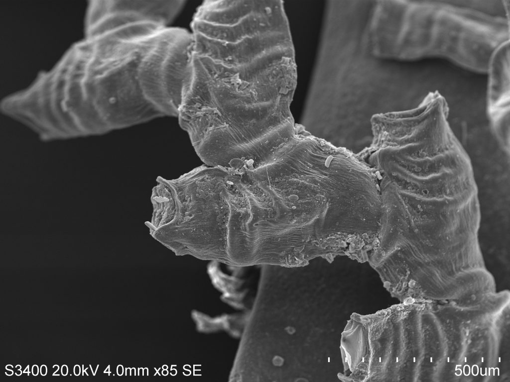
These SEM photos below show the chitonous exoskelton, or perisarc, of the colonial hydroid Obelia. This species is well-studied by scientists and is commonly used in zoology classes to describe the basic hydroid body plan and life cycle. The main body of the colony is composed of living tissue known as the coenosarc, which is covered by the perisarc. Colonies can grow in a variety of patterns, which are often used for identification purposes. Obelia, for example, demonstrates the branching pattern some hydroids adopt, while others grow vertically off of a common stolon.


Both body forms indicative of the greater Cniderian grouping, the medusa and the polyp, are present in this species. Medusae are released from the gonozooids, producing free-swimming medusae velum with gonads, a mouth, and tentacles. The medusae reproduce sexually, releasing sperm and eggs that fertilize to form a zygote, which later morphs into a blastula, then a ciliated swimming larva called a planula. The planulae live as free-swimming members of the planktonic community before eventually attaching themselves to a solid surface, where they begin their reproductive phase of life. Once attached to a substrate, a planula quickly develops into one feeding polyp. As the polyp grows, it begins developing branches of other feeding individuals, thus forming a new generation of polyps by asexual budding.


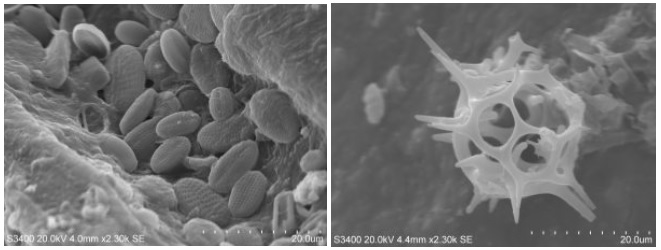
If you read Elizabeth's recent post regarding crustose coralline algae, you saw that it's very common to find unexpected organisms associated with your target species. As I was imaging this hydroid sample, I came across a few stow-aways that were too interesting not to include. On the left is a cluster of diatoms, marine phytoplankton responsible for much of the primary production in our oceans. They are often used in analyzing sediments, as discussed in some of Jennifer's posts in this atlas. On the right is an unidentified planktonic organism. Even when it's not possible to ID something on site, we can use these images to make inferences regarding their life history. For example, spines such as these are thought to function in decreasing the rate of sinkage for these organisms. At such small sizes (not the scale), water represents a very viscous medium, and increasing surface area is one way to create drag and stay afloat.


The inside scoop on Giant Kelp

If you live by the ocean, especially on the California coast, then you are probably familiar with Giant Kelp. This is the species of kelp that creates large brown forests underwater and lovely fly infested lumps of smelly nastiness on the beaches. Giant Kelp, known to phycologists as Macrocystis pyrifera, is one of the most common seaweeds people associate with the ocean. This is because this particular species is found year round. Unlike other seaweeds that tend to have seasonal debuts, Giant Kelp has a special mechanism for growth and reproduces year round, making it resistant to seasonal changes in its environment.

Giant Kelp is a type of algae, not a type of plant like what you would think of on land. Land plants grow using xylem and phloem, two mechanisms responsible for storing water and transferring nutrients throughout the structure, respectively. Algal species are not related to land plants and therefore do not have these same mechanisms for growth. Instead, in algae like kelps, they have what is called trumpet hyphae, which look like a bunch of trumpets that move nutrients throughout the entire thallus. This mechanism is extremely successful. In fact, the Giant Kelp species is so successful at transferring its nutrients that it can, in the best environmental conditions, grow up to a foot a day! This rapid growth helps keep Giant Kelp around during any season, creating the forests that we all know and love.
Giant Kelp is also resistant to seasonality due to its reproductive methods. This particular species has what we call sporophylls which are specialized blades specifically grown to reproduce. Sporophylls are full of reproductive spores and they reside at the bottom of the frond just above the holdfast. It is most obvious that they are ready to reproduce when the blades turn a milky white. For this particular segment I wanted to use the Scanning Electron Microscope to look at the cross section of sporophyll blade. I cut a small square out of the milky sporophyll blade I had collected and stuck it on a stub.
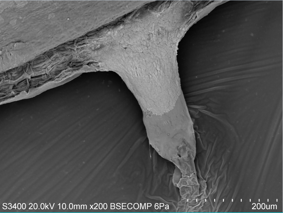
You can see in the picture to the left that you can view the top and bottom of the blade but the focus is on the inside. You can see the honeycomb structure inside the blade. When I scanned over to this part of the cross section I found this odd structure oozing out of my cross section. After chatting with a few of my lab mates we have decided that the sporophyll was oozing out some sugar mucilage. This means that this particular blade was full of good nutrients!


The more I work with algae the more I realize how complicated it is to work with tissue on the SEM instead of hard organic material like some of my class mates. I find it as a fun challenge to continue to understand the microscopic structures of algae!

Coralline Algae…. the unsung hero

Many I am sure have seen the beautiful pink and purple hues that dance in the tide pools during low tide. Some may have also seen pink or purple looking rocks in the sand along Monterey Bay's coast.

These beautiful colors you see are not a kind of coral or a type of rock, contrary to common belief. Their name can be misleading but corallines, crustose and articulated, or upright and branched, are actually types of red algae found in many habitats around the world. They get their name because of the calcium carbonate within their cell structures on their thallus that creates a hard outer form of protection from the environment and hungry grazers. An articulated coralline can be found in the low intertidal and the subtidal. Its hard structure provides protection from breakage when living in areas like the low intertidal where wave energy can be high. Because the structure is not completely rigid, there is just enough flexibility for it to sway back and forth with the tides. The calcium carbonate within their thallus also provides protection from grazing invertebrates. Instead of having a fleshy outer structure, the coralline has a hard rough structure that doesn't seem as appetizing. However, there are a few choice grazers who actually prefer coralline, especially in its crustose form, like chitins that have a rough tongue called a radula that is used to scrape food off of rocks. 

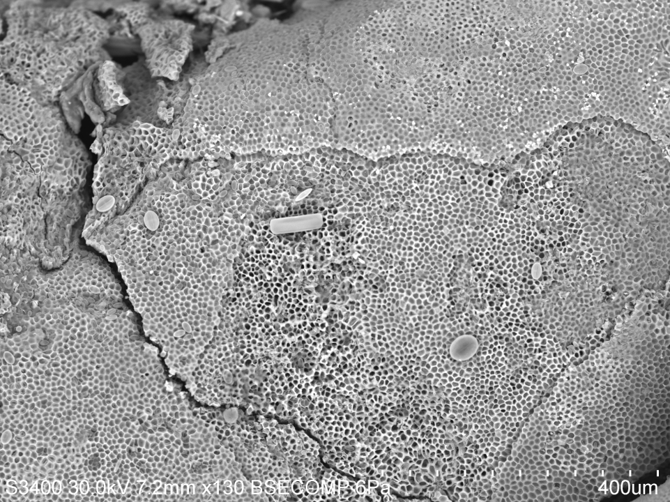
If you look at this SEM photograph to above you can see the calcium carbonate structure well. What you are seeing are individual layers of cells on the outer most crustose shell shedding off. Some species of coralline algae do this to anti-foul or get rid of any epiphytes that may be eating away and hindering the health of the individual. This was first seen using scanning electron microscopy methods and as research continues to evolve, more studies are finding that this is mechanism is quite common especially in crustose species of coralline.

If you look even closer, like at the SEM picture below, you can see the honeycomb structure of the cells. These cells have that hard structure made of calcium carbonate, also known as limestone. What is truly fascinating about coralline algae, espeically the crustose form, is that it is actually an unsung hero for the coral reefs in tropical environments. When the crustose coralline algae settles and starts to grow, it creates a glue or cement that ultimately keeps coral reef beds together. Because of its limestone cellular structure, coralline has been shown to positively influence coral reefs worldwide. If you want to learn more, follow this link.

I purposely chose to not wash off the coralline very well before creating a stub so that I could see what kinds of things may have been living on my articulated coralline specimen.
In this picture to the right you can see there are a few geometric shapes on top of the structure. These are all different kinds of diatoms that were either just in the water that was brought in with the coralline or they were living on top of the actual structure. The most exciting thing about using the scanning electron microscope is finding what other microscopic things may be living with your species of interest.

Minerals in the Sand

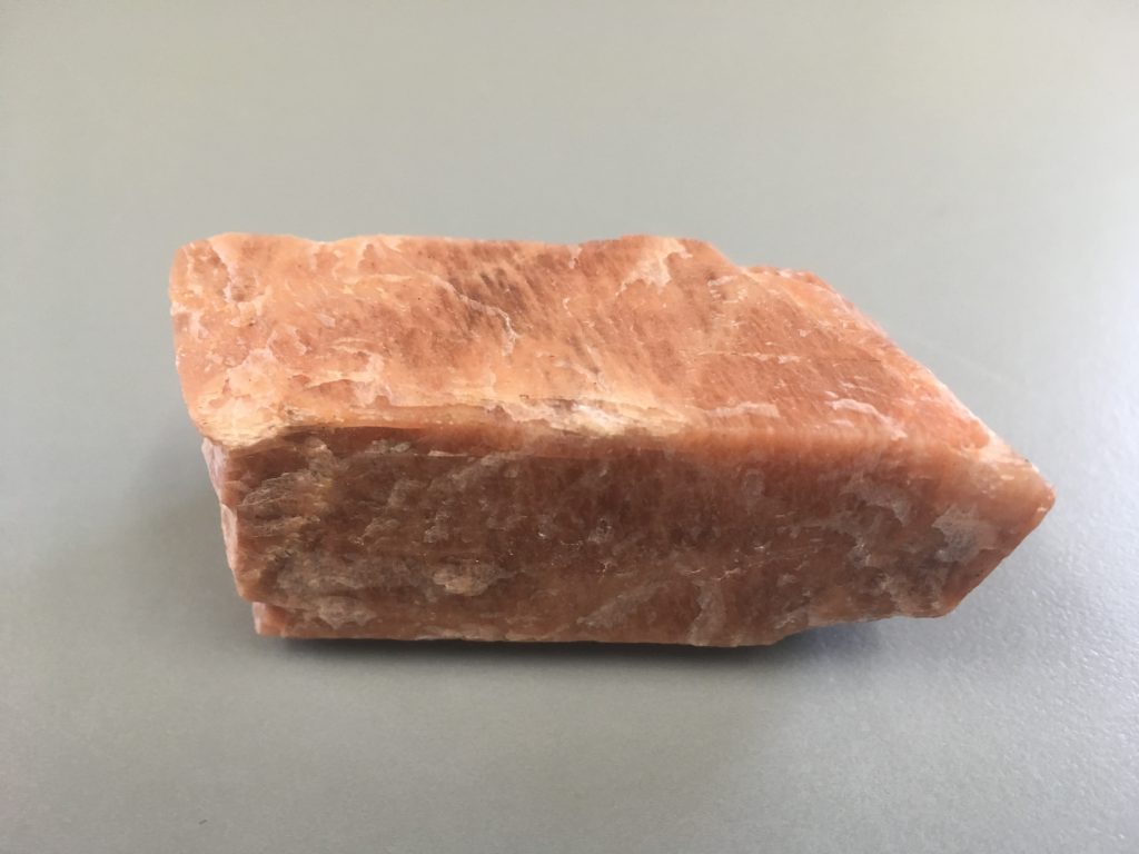
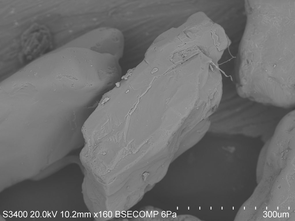
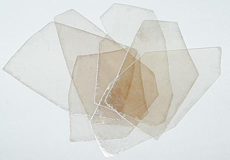
Feldspars: Feldspars are the most abundant minerals in the Earth’s crust. This group of silicate minerals is somewhat hard (6-6.5 on Moh’s hardness scale), is often pink, white, or grey in color, and has two good cleavage planes that meet at nearly a 90° angle (figure 1). There are two main types of feldspar, potassium (KAlSi3O8) and plagioclase (NaAlSi3O8 or CaAl2Si2O8). Plagioclase is further divided by the relative amount of sodium and calcium within the mineral. Figure 2 and 3 shows SEM images of these minerals within beach sand.

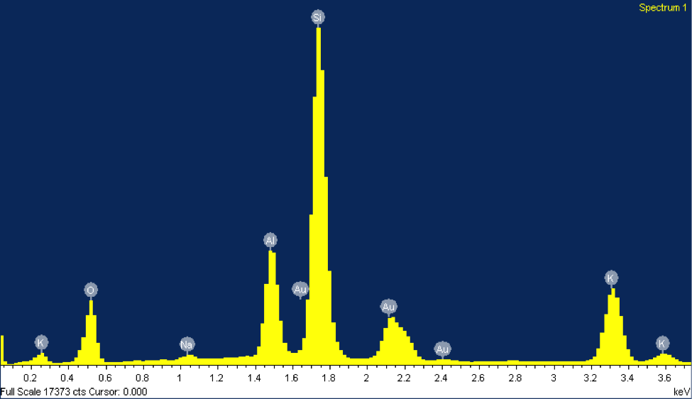
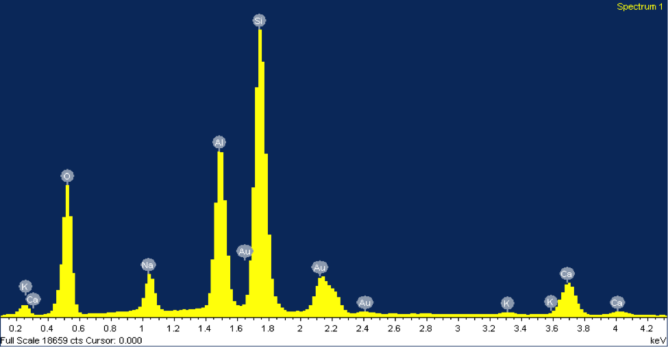
The EDX elemental spectrum for each mineral shows the abundance of various elements detected, the main tool used to identify these grains. In EDX analysis, inner shell electrons of atoms composing the mineral are excited and ejected. Outer shell electrons replace these ejected electrons to maintain stability, releasing specific amounts of x-ray energy corresponding with a particular element. This energy is shown on the x-axis of the EDX elemental spectrum. The minerals in figures 2 and 3 both contain peaks of silicon. Notice how the peaks align at the same energy level for both minerals, about 1.74 keV. The y-axis represents the counts of each element—the higher the count, the more abundant that element is within the mineral. However, peaks of gold can be disregarded since the sample was coated with gold. The mineral in figure 2 was identified as potassium feldspar due to peaks in silicon, aluminum, and potassium.

The counts suggest that silicon is about three times more abundant than aluminum and potassium, matching its chemical formula, KAlSi3O8. The mineral in figure 3 was identified as plagioclase feldspar due to the presence of silicon, sodium, calcium, and aluminum at abundances matching its chemical formula, NaAlSi3O8 or CaAl2Si2O8. Often plagioclase feldspar is referred to on a spectrum from sodium-rich to calcium-rich. Our specimen falls somewhere in the middle, with almost equal parts sodium and calcium. Morphologically, the grain of plagioclase feldspar displays the classic cleavage often associated with feldspars, with the flat surface on top.


Figure 1: (Right) Potassium felpspar and (left) plagioclase feldspar.  

Figure 2: (Right) A SEM image of a grain of potassium feldspar from beach sand collected at Moss Landing Beach. (Left) The EDX elemental spectrum from the feldspar grain.

Figure 3: (Right) A SEM image of a grain of plagioclase feldspar from beach sand collected at Moss Landing Beach. (Left) The EDX elemental spectrum from the feldspar grain.

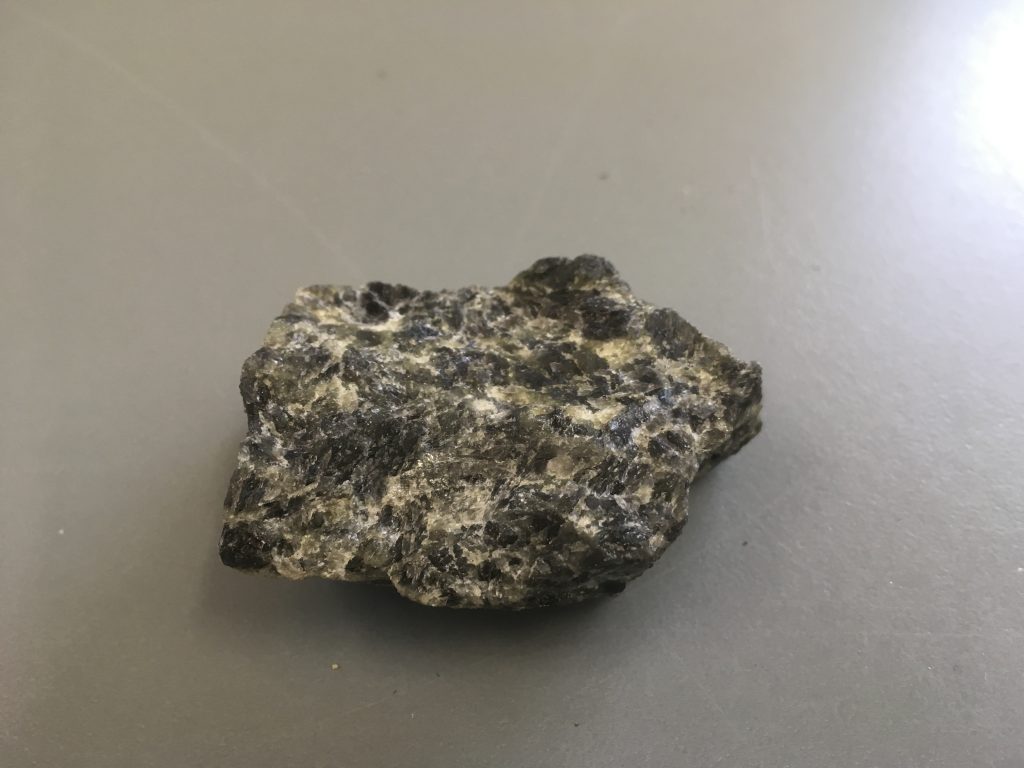
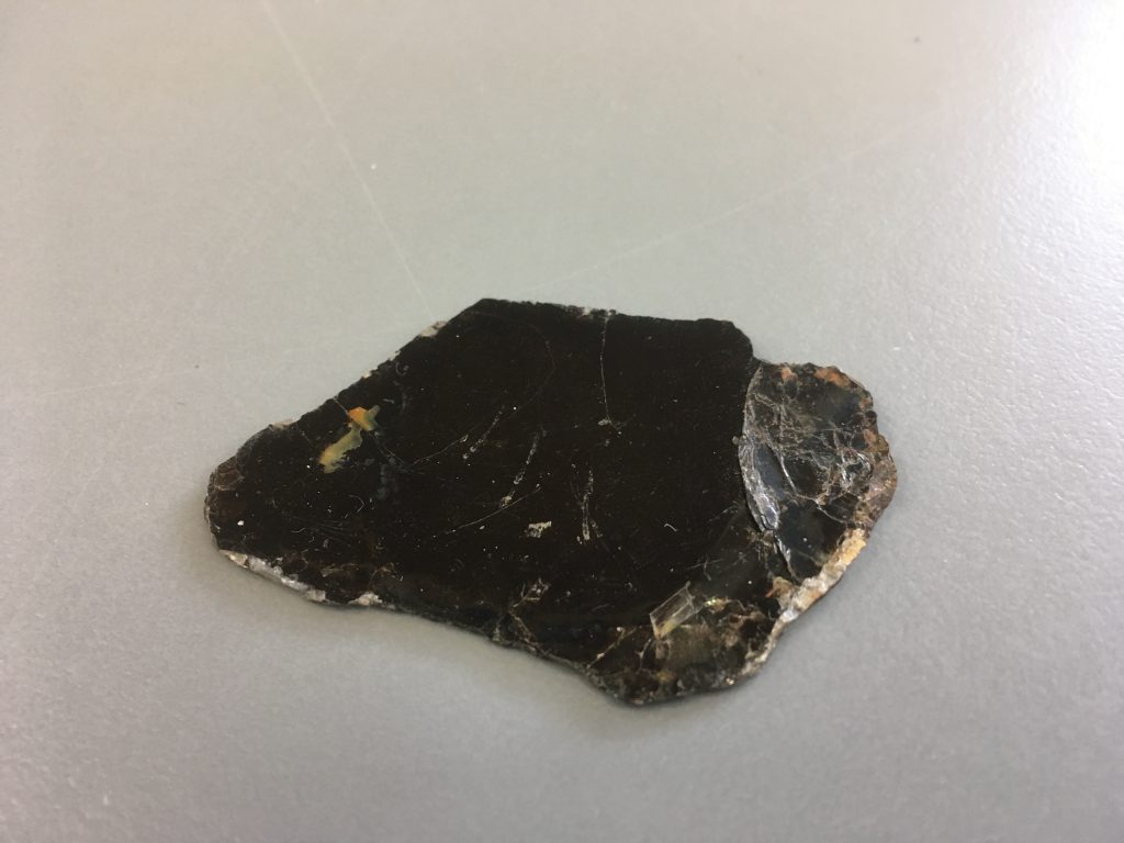
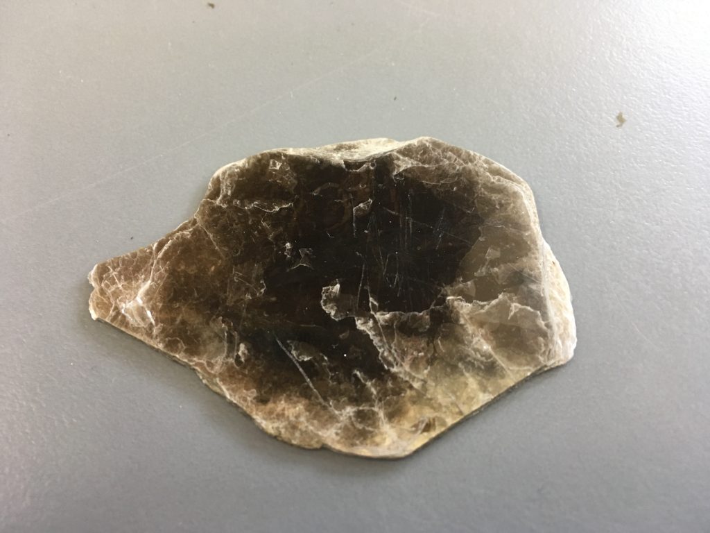
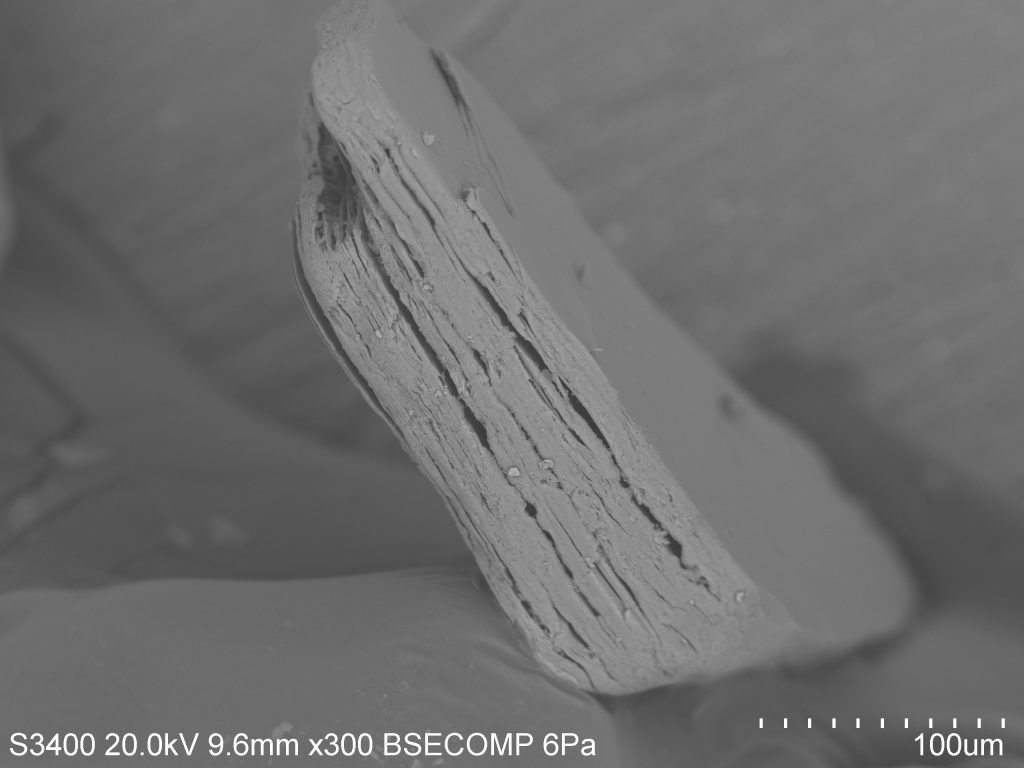
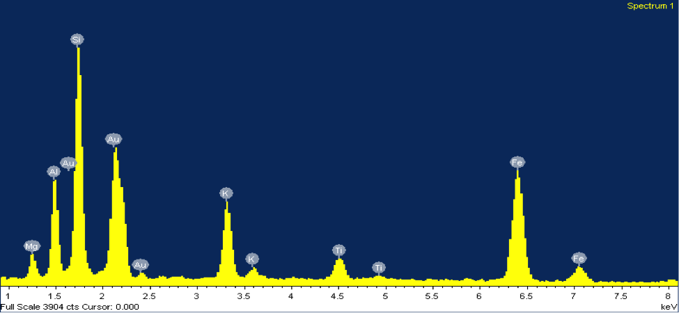
Mica: Biotite mica is a magnesium and iron rich mineral, usually black in color and quite soft (2.5-3 on Moh’s hardness scale). Biotite shares many properties with the potassium-rich muscovite mica, which is usually much lighter in color due to a lack of magnesium/iron (figure 4). The most prominent of these properties is the sheet-like formation of micas (one sheet is about 0.003-0.1 millimeters thick). Figure 5 shows an SEM image of a grain of biotite from beach sand collected at Moss Landing Beach. When I first came upon this grain under the SEM I knew that it was mica, due to its sheet-like form and good cleavage in one direction. However, from the SEM image there is no way of knowing whether it is muscovite or biotite—so I turn to the EDX. The EDX elemental spectrum (figure 5) clearly shows a significant presence of magnesium and iron, indicating that this grain is biotite mica.

Figure 4: (Right) Biotite mica and (left) muscovite mica. The color difference in these two similar minerals is due to the presence of magnesium/iron in biotite. Notice the sheet-like form of both minerals.  

Figure 5: (Right) SEM image and (left) EDX elemental spectrum of a grain of biotite mica from beach sand collected at Moss Landing Beach.  

What to think about when stung by a stingray

The tip of the barb is the sharpest part of the structure and allows the animal to impale predators.

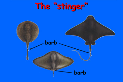
Although some ray species do not have stingers there is a large portion of the group that have them. These structures are not used to hunt for prey but rather as a way to defend themselves from predators. Occasionally people have interactions where they are stabbed by them and it can be a serious situation.


The manta ray (left) is a species that is well known and is absent of a barb. The barb is commonly located at the base or close to the base of the tail (right)

Back in April of 2015 I had an interaction with a bat ray where I was stung in the arm. The initial pain was brief and my adrenaline began to kick in. I asked my friend to take the barb out immediately and thinking about it I may have wanted to keep it in. When thinking about the barb it’s in the shape of a Christmas tree almost. The barb has a sharp point in the top and it’s followed by a series of serrations facing the opposite side of the initial point and taking it out could do more damage than leaving it in, but I was in survival mode and I wanted it out of my body.


This is an image showing the anatomy barbs come in. The darker sections labeled with the number 1 is where the venom glands are located. The number 2

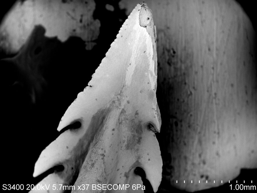
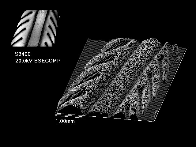
For this post I wanted to look at a barb under the SEM from the same species that stung me and get an appreciation for this defense mechanism. As seen in the second and third picture there are variations in shape and size and it helps with species identification if that is wall that you have to work with. In my ichthyology class we dissected bat rays, where I collected one of the barbs. I also used the Oblique analysis tool to get a three dimensional image of the barb. Can you tell where the venom gland would be located on the barb in relation to picture 3?


These are the images of the barb that specifically focuses on the serrations. The serrations on this structure help the ray increase its capacity of damage if the barb is pulled out.


Stepping into the ring(s)

On the first day of our Scanning Electron Microscopy class our instructor asked if we would like to prepare a sample for the following week. I initially didn’t have a clue on what I wanted to use because I had not prepared any sample of my own to go through the process to be prepped for the SEM. While I was waiting and thinking about what I wanted to use, I realized that I had a pair of otoliths in my backpack. These otoliths were collected from a rock fish during a series NOAA surveys conducted off the West Coast of the U.S. I had them as mementos of the trip I was on and forgot about them for a couple of months and I thought this would be a great opportunity to look at one of them under a high powered microscope.

Otoliths are boney structures located behind the brain of fish and come in different shapes and sizes. Otoliths are not found in cartilaginous fish like sharks, skates, rays and chimaeras and are predominantly in bony fishes. These hard structures are made of calcium carbonate and aid fish in detecting balancing, and directional movement. Scientist can use these structures to identify species, calculate growth rates, and determine the age of the fish. This is made possible because during the lifespan of these fish proteins and calcium carbonate layer on top of each other annually.

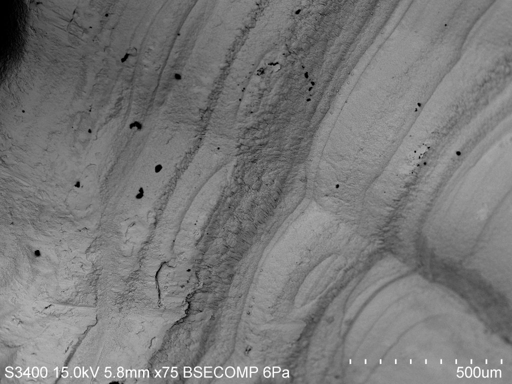
(Picture 1) A comparison in size of an otolith and a penny. Although it’s coated in a thin layer of gold you can see the distinct bands that surround the otolith. These distinct dark “rings” are made of calcium carbonate and proteins, are commonly used to age fishes. This technique is analogous to counting the rings of a tree to determine the age but I was curious to see if there was physical difference between the bands or if it is just a difference in color. When we actually looked at the otolith it almost looked like the surface of a planet. Some areas of the otolith looked like canyons and it was interesting seeing how distinct the bands looked compared to the lighter areas of the otolith. When we looked even closer it seemed that there was a courser texture in the bands than areas without it and the structures were smaller. I thought it was an interesting thing to see not only that there is a color difference but a physical difference in a micro scale.

(Picture 2) A first glance at the surface of the otolith where distinct banding paterns are easily seen. The box is a lead-in to pictures 3&4 where we get a deeper understandig of the physical aspects of the bands.

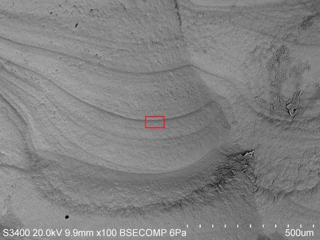
(Picture 3) The physical differences between the darker band and the lighter bands. The red box leads into the physical aspect of the smaller calcium carbonate structures in Picture 4.

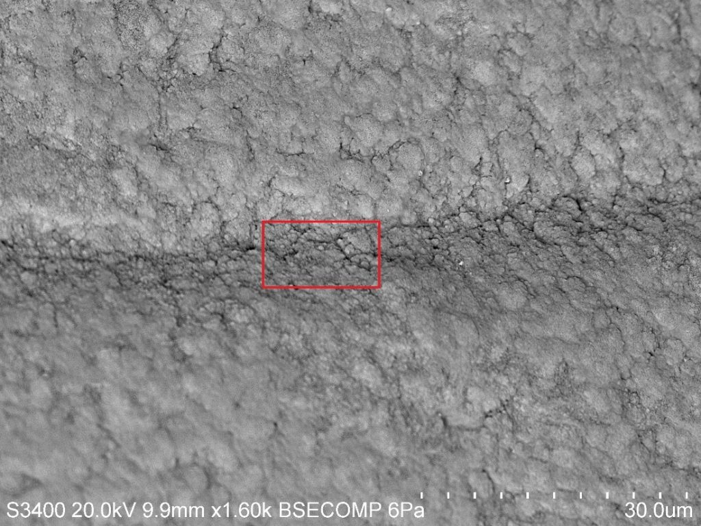
(Picture 4) The structures in the band almost mirror the structures on the light side but are smaller and more compact.

Sea-Urchin’ for Detail at a Micro Level

Echinoderms are an interesting bunch of critters, encompassing intertidal favorites such as the star fish, the sea cucumber, and the sea urchin. The roots of the word "Echinoderm" stem from the Green word "ekhinos," meaning porcupine or hedgehog, and the root -derm, meaning skin. In my opinion, sea urchins represent the epitome of a spiky-skinned animal, and in light of that fact I thought I'd look at a few spines under the SEM.

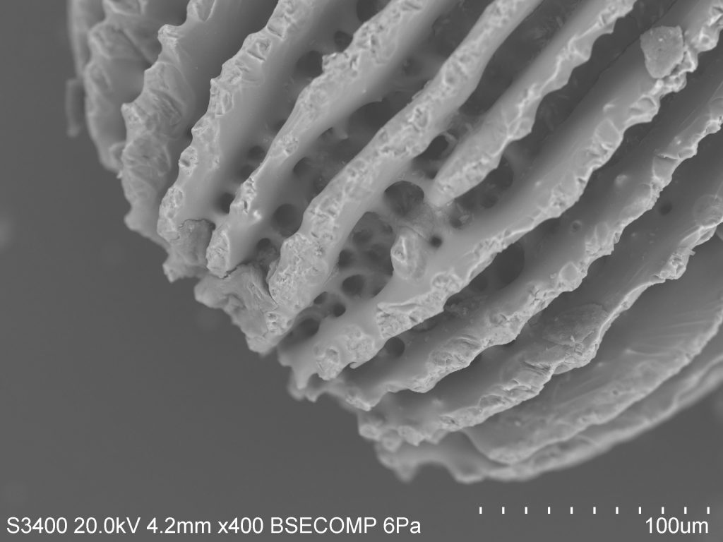
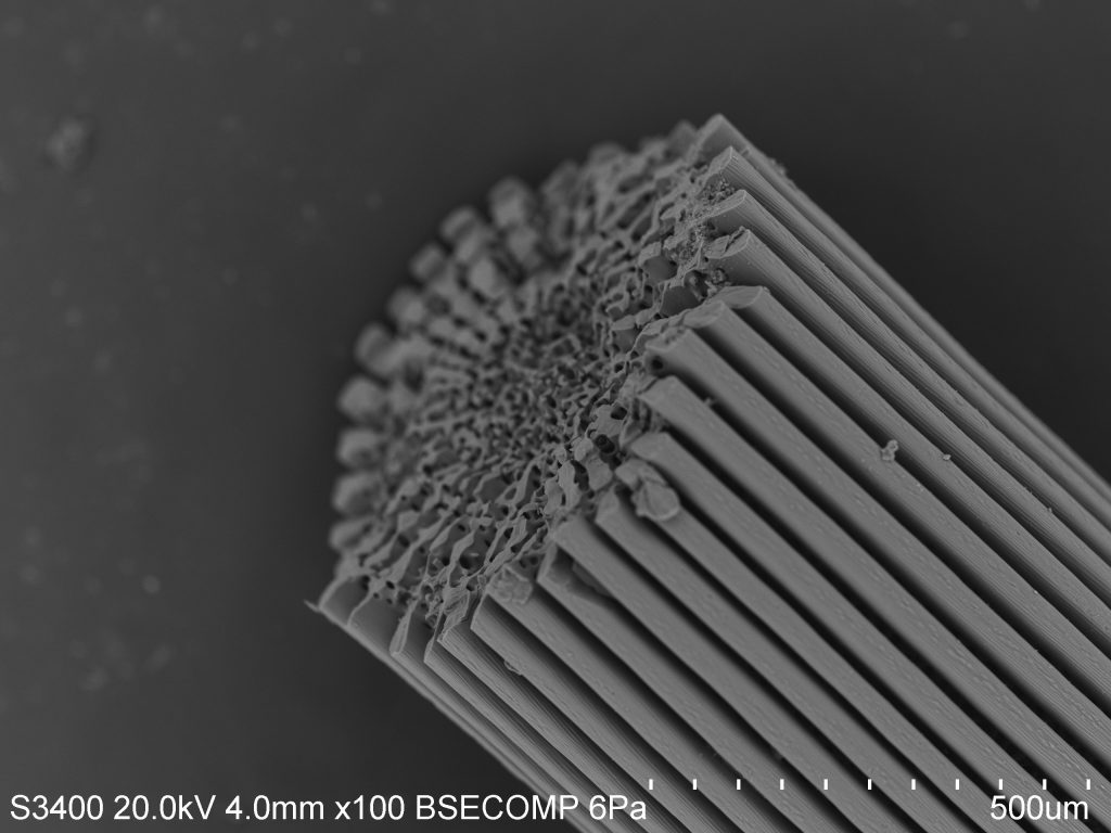
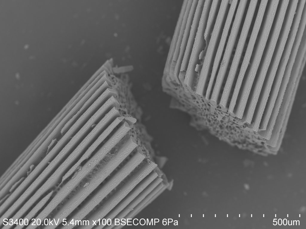
As you can see, the spines are ridged and porous. Sea urchins do not have a closed circulatory system and instead rely on an open water vascular system to help with the uptake of nutrients, the flushing out of wastes, locomotion, and respiration. As such, much of their body plan is open to the environment. In this case, it is actually covered by a thin layer of epidermis (the outer layer of cells), making the spines themselves a feature of the interior endoskeleton. You can think of this as similar to the skin that covers our bones, although spines and bones serve very different purposes.


The name for this internal structural organization is the stereom. The stereom is a feature that all echinoderms share, and is useful in identifying lineages in fossilized samples. Spines mainly show two distinct morphological configurations: the base, made of a meshwork stereom, and the shaft, which has several longitudinal septa and a central core of meshwork stereom. This can be seen distinctly in the photo. The stereom is sponge-like and varies in composition by species. In a sea urchin, the stereom may be 50% living cells such as connective tissue cells and phagocytes involved in nonspecific defense. The rest will be composed of a matrix of calcite sclerocytes which direct mineral production and repair.

Over several days, sea urchins are actually able to regrow spines in places where damage has been inflicted, or to grow entirely new spines. The urchins begin growing micro-spines in a conical shape by precipitating calcite from the surrounding seawater. As these small spines thicken, lateral growth takes place, allowing neighboring micro-spines to join in a horizontal "bridge." The formation of bridges results in the mesh of cells spanning from the base of the spines to the tip.


All this terminology is only the tip of the iceberg (tip of the spine?), and I'm keen to look further. The test of the sea urchin, which is the "hard shell" under the spines and feet and skin, is full of microscopic pores through which several accessory structures pass. The geometry is quite interesting from a macro-perspective, as all echinoderms share pentaradial symmetry, meaning their bodies are grouped loosely into five identical parts. In urchins, these manifest as alternating ambulacral (related to the feet) and inter-ambulacral (between the feet) regions. I imagine the micro-analysis of differences between these regions would be interesting as well.


Piston Core 36

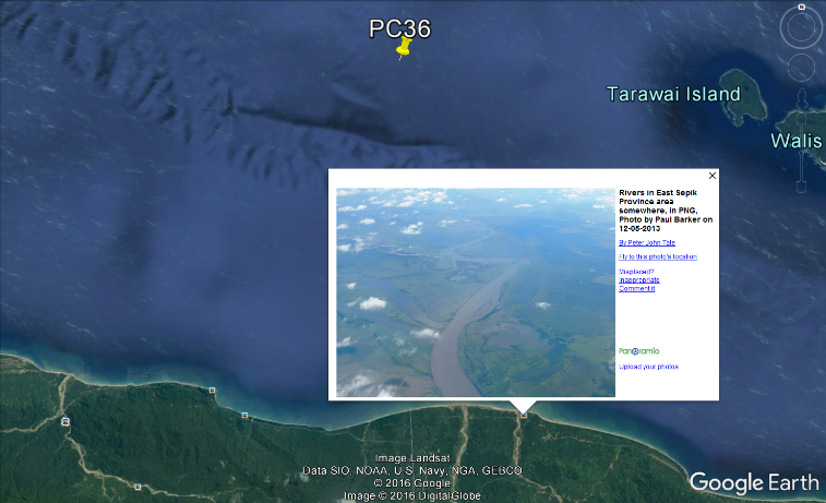
In previous posts we have discussed deep-sea sediments that are rich in skeletal debris. Now we will focus on a site that is abundant in siliciclastic material. Piston core 36 (PC36) is a site located adjacent to the Western Sepik River at a water depth of 903 m. The sample we will be focusing on was collected at a subsurface depth of 139-140 cm. Its close proximity to the delta may the source of such terrigenous material.


The amount of sediment a river can transport changes over time, therefore, sediments found at varying depths can reveal changes in precipitation. Change in precipitation is a result of climate change, and as climate changes so do the environments that are in the region! A comparison of siliciclastic material will be addressed in an upcoming post where we will look at another sample from PC36, but at a subsurface depth of 218-219 cm. Siliciclastic rocks are non-carbonate sedimentary rocks that are almost exclusively silica bearing. They are terrigenous material, derived from erosion or weathering of land-based rocks, transported by wind or water. Common siliciclastic minerals are quartz, feldspars, micas, and heavy element minerals.


The images above highlight a common sheet silicate mineral, known as a mica. Its platy texture is easily distinguishable on the adjacent minerals in the lower and upper left corners. This platy texture is the result of basal cleavage; minerals in the mica group have one cleavage plane and can be peeled into perfect thin sheets. Mica formations are generally associated with volcanoes and hydrothermal vents. Several of the brighter minerals scattered across the surface of the mica are calcite (CaCO3) crystals.


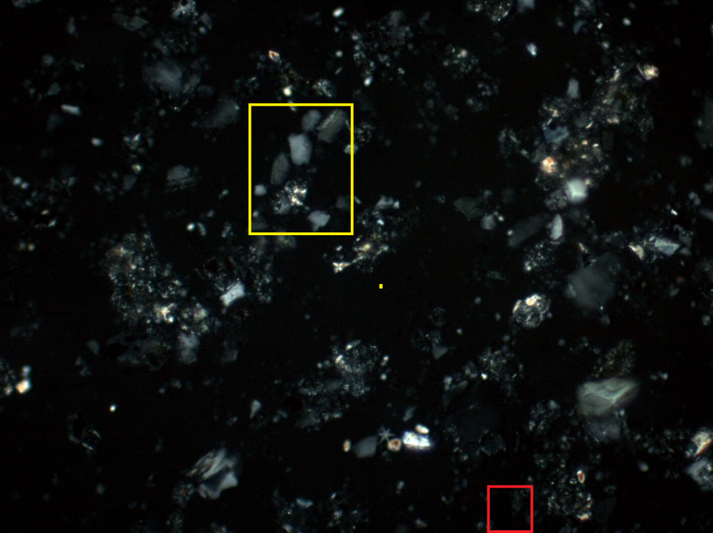
As expected the sediment from PC36 is highly siliciclastic (yellow), but contains very few quartz crystals, in PPL (right) and XPL (left).  Samples containing quartz are very stable and mature sediments. Therefore, this sample appears to be freshly deposited given its immature mineral assemblage. This supports the hypothesis that this sediment may have been transported from land by the Sepik River. This sample also contains a fair amount of opaque (red) minerals. Opaque minerals prevent light from passing through and commonly contain iron oxides and sulpihides. It is apparent that this sediment contains minimal skeletal debris as compared to previous sediments we have looked at. For comparison, see the previous post regarding PC23.