Coralline Algae…. the unsung hero

Many I am sure have seen the beautiful pink and purple hues that dance in the tide pools during low tide. Some may have also seen pink or purple looking rocks in the sand along Monterey Bay's coast.

These beautiful colors you see are not a kind of coral or a type of rock, contrary to common belief. Their name can be misleading but corallines, crustose and articulated, or upright and branched, are actually types of red algae found in many habitats around the world. They get their name because of the calcium carbonate within their cell structures on their thallus that creates a hard outer form of protection from the environment and hungry grazers. An articulated coralline can be found in the low intertidal and the subtidal. Its hard structure provides protection from breakage when living in areas like the low intertidal where wave energy can be high. Because the structure is not completely rigid, there is just enough flexibility for it to sway back and forth with the tides. The calcium carbonate within their thallus also provides protection from grazing invertebrates. Instead of having a fleshy outer structure, the coralline has a hard rough structure that doesn't seem as appetizing. However, there are a few choice grazers who actually prefer coralline, especially in its crustose form, like chitins that have a rough tongue called a radula that is used to scrape food off of rocks. 

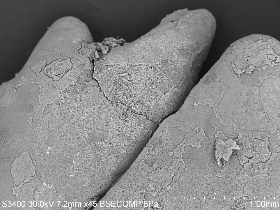
If you look at this SEM photograph to above you can see the calcium carbonate structure well. What you are seeing are individual layers of cells on the outer most crustose shell shedding off. Some species of coralline algae do this to anti-foul or get rid of any epiphytes that may be eating away and hindering the health of the individual. This was first seen using scanning electron microscopy methods and as research continues to evolve, more studies are finding that this is mechanism is quite common especially in crustose species of coralline.

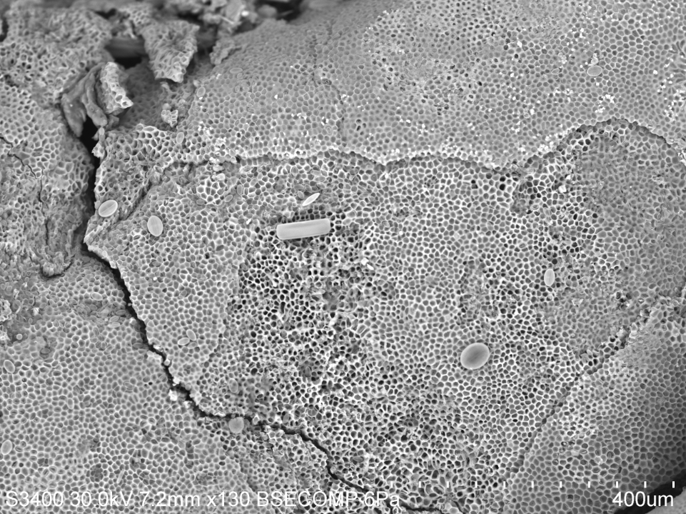
If you look even closer, like at the SEM picture below, you can see the honeycomb structure of the cells. These cells have that hard structure made of calcium carbonate, also known as limestone. What is truly fascinating about coralline algae, espeically the crustose form, is that it is actually an unsung hero for the coral reefs in tropical environments. When the crustose coralline algae settles and starts to grow, it creates a glue or cement that ultimately keeps coral reef beds together. Because of its limestone cellular structure, coralline has been shown to positively influence coral reefs worldwide. If you want to learn more, follow this link.

I purposely chose to not wash off the coralline very well before creating a stub so that I could see what kinds of things may have been living on my articulated coralline specimen.
In this picture to the right you can see there are a few geometric shapes on top of the structure. These are all different kinds of diatoms that were either just in the water that was brought in with the coralline or they were living on top of the actual structure. The most exciting thing about using the scanning electron microscope is finding what other microscopic things may be living with your species of interest.

Minerals in the Sand

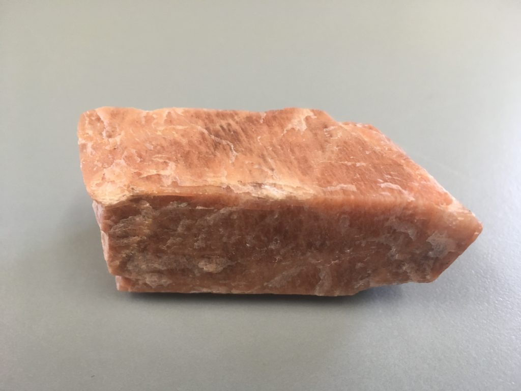
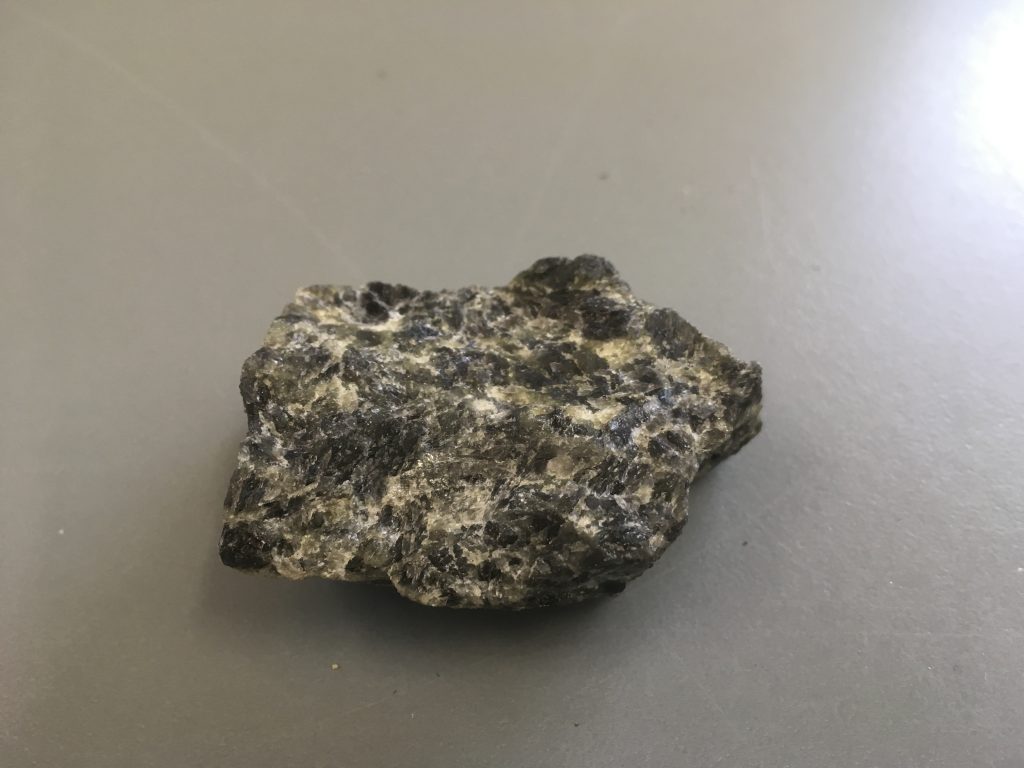
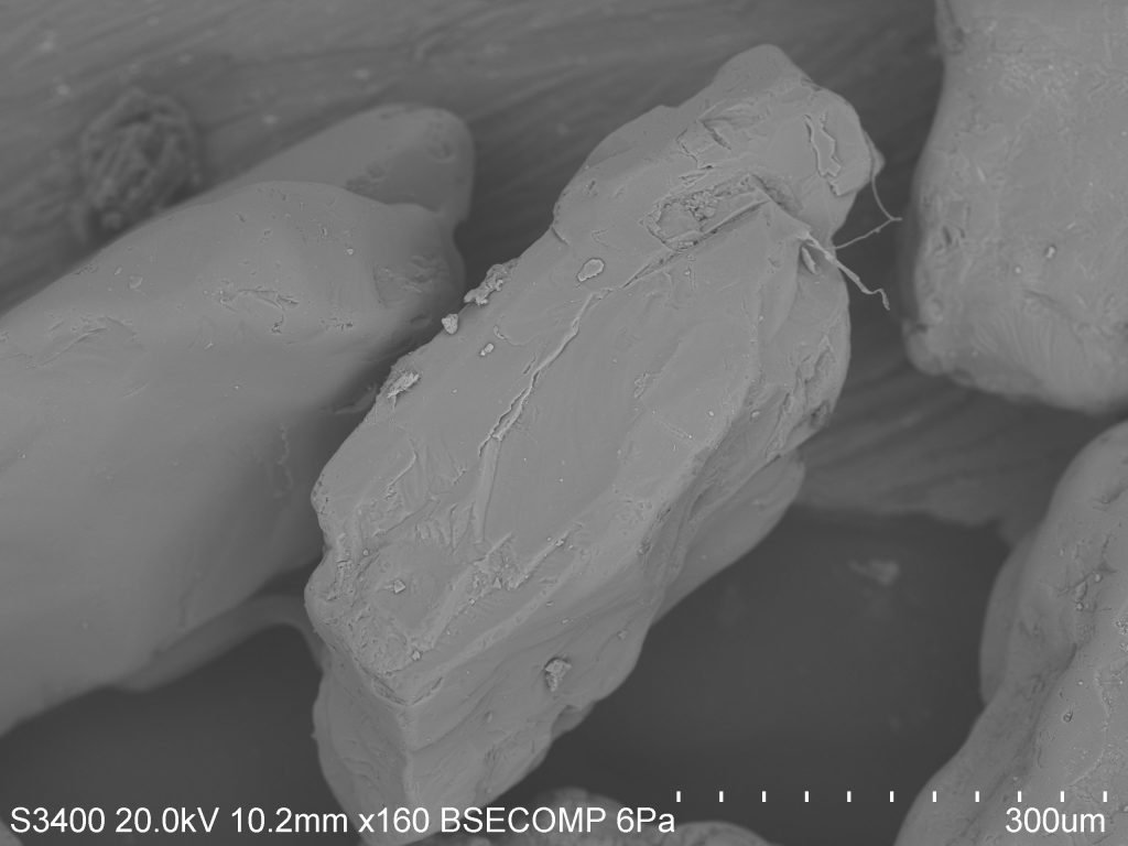
Feldspars: Feldspars are the most abundant minerals in the Earth’s crust. This group of silicate minerals is somewhat hard (6-6.5 on Moh’s hardness scale), is often pink, white, or grey in color, and has two good cleavage planes that meet at nearly a 90° angle (figure 1). There are two main types of feldspar, potassium (KAlSi3O8) and plagioclase (NaAlSi3O8 or CaAl2Si2O8). Plagioclase is further divided by the relative amount of sodium and calcium within the mineral. Figure 2 and 3 shows SEM images of these minerals within beach sand.

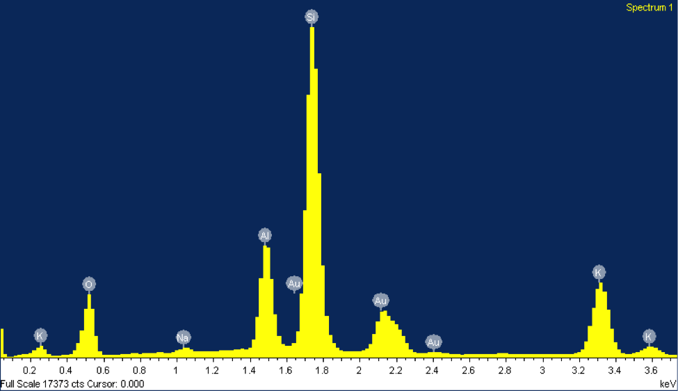
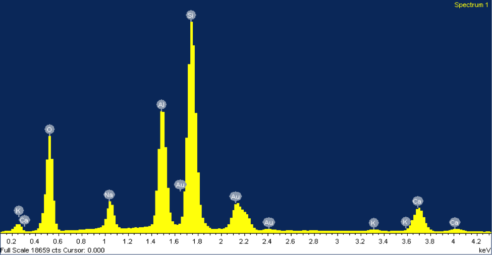
The EDX elemental spectrum for each mineral shows the abundance of various elements detected, the main tool used to identify these grains. In EDX analysis, inner shell electrons of atoms composing the mineral are excited and ejected. Outer shell electrons replace these ejected electrons to maintain stability, releasing specific amounts of x-ray energy corresponding with a particular element. This energy is shown on the x-axis of the EDX elemental spectrum. The minerals in figures 2 and 3 both contain peaks of silicon. Notice how the peaks align at the same energy level for both minerals, about 1.74 keV. The y-axis represents the counts of each element—the higher the count, the more abundant that element is within the mineral. However, peaks of gold can be disregarded since the sample was coated with gold. The mineral in figure 2 was identified as potassium feldspar due to peaks in silicon, aluminum, and potassium.

The counts suggest that silicon is about three times more abundant than aluminum and potassium, matching its chemical formula, KAlSi3O8. The mineral in figure 3 was identified as plagioclase feldspar due to the presence of silicon, sodium, calcium, and aluminum at abundances matching its chemical formula, NaAlSi3O8 or CaAl2Si2O8. Often plagioclase feldspar is referred to on a spectrum from sodium-rich to calcium-rich. Our specimen falls somewhere in the middle, with almost equal parts sodium and calcium. Morphologically, the grain of plagioclase feldspar displays the classic cleavage often associated with feldspars, with the flat surface on top.


Figure 1: (Right) Potassium felpspar and (left) plagioclase feldspar.  

Figure 2: (Right) A SEM image of a grain of potassium feldspar from beach sand collected at Moss Landing Beach. (Left) The EDX elemental spectrum from the feldspar grain.

Figure 3: (Right) A SEM image of a grain of plagioclase feldspar from beach sand collected at Moss Landing Beach. (Left) The EDX elemental spectrum from the feldspar grain.

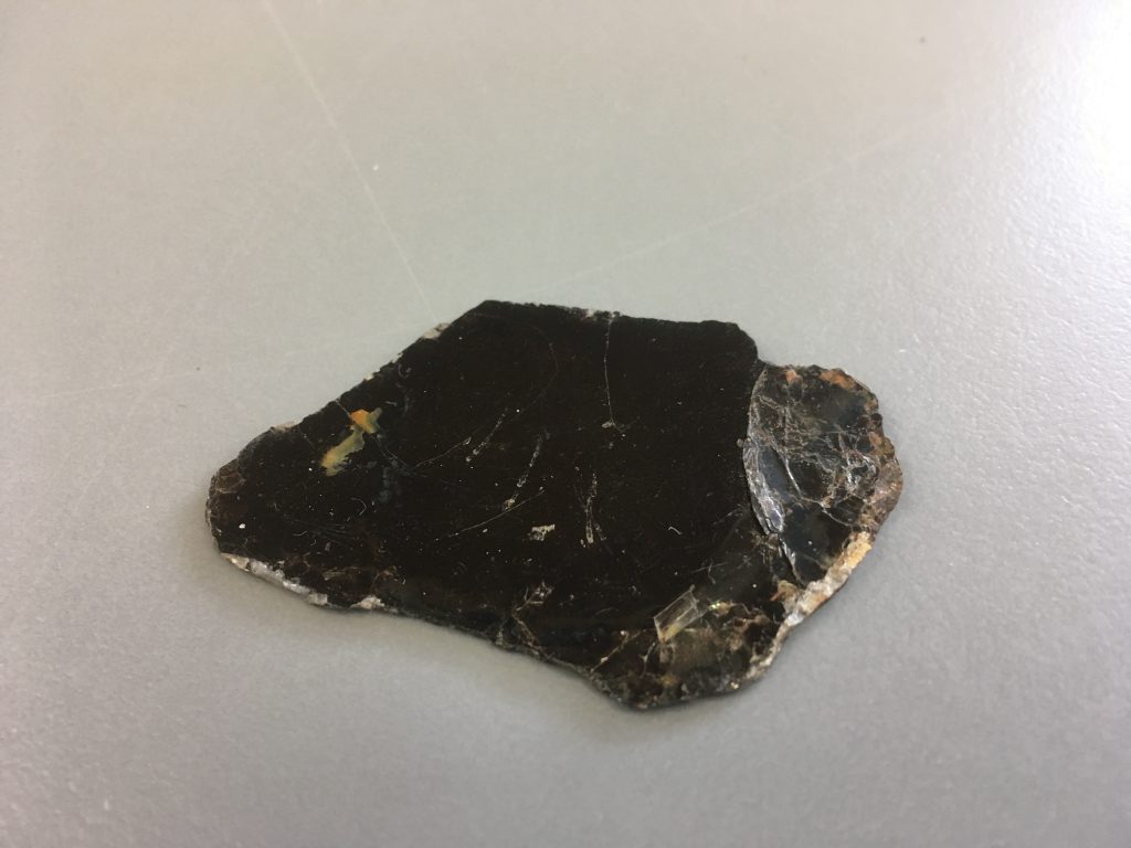
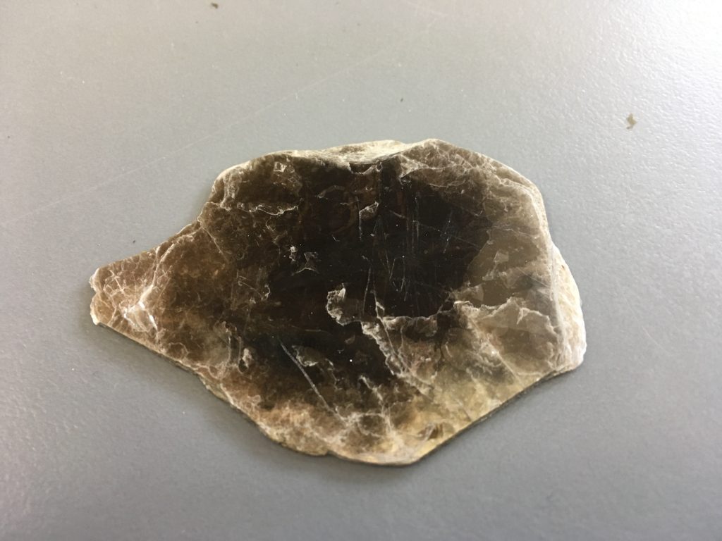
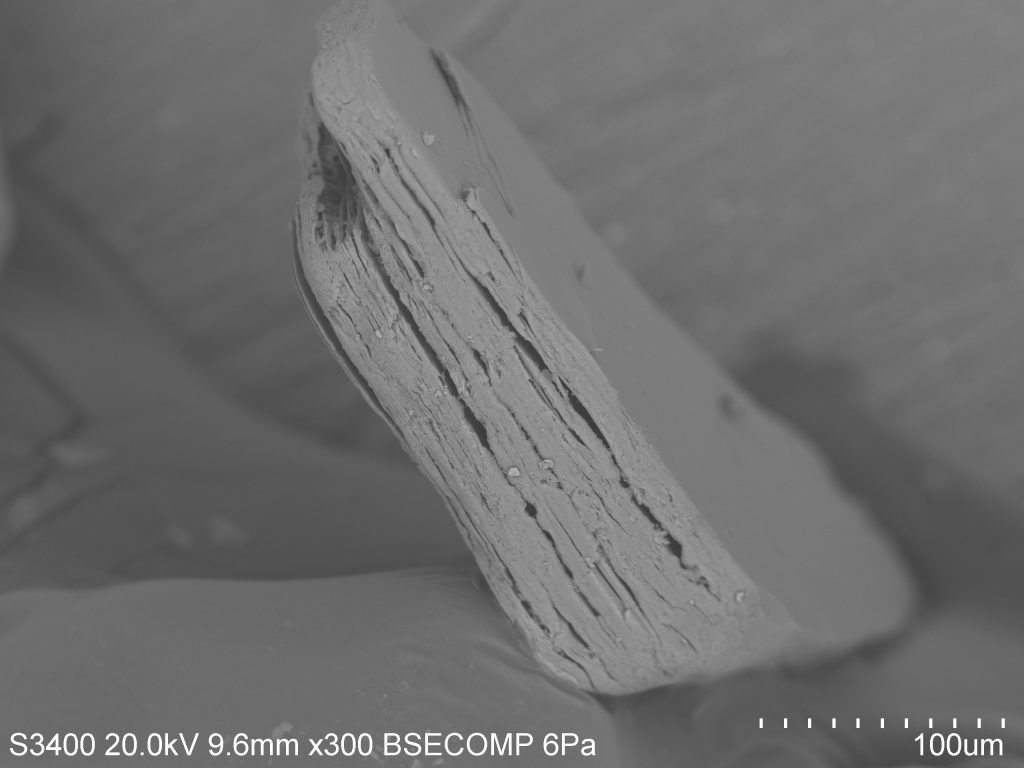
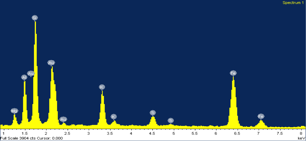
Mica: Biotite mica is a magnesium and iron rich mineral, usually black in color and quite soft (2.5-3 on Moh’s hardness scale). Biotite shares many properties with the potassium-rich muscovite mica, which is usually much lighter in color due to a lack of magnesium/iron (figure 4). The most prominent of these properties is the sheet-like formation of micas (one sheet is about 0.003-0.1 millimeters thick). Figure 5 shows an SEM image of a grain of biotite from beach sand collected at Moss Landing Beach. When I first came upon this grain under the SEM I knew that it was mica, due to its sheet-like form and good cleavage in one direction. However, from the SEM image there is no way of knowing whether it is muscovite or biotite—so I turn to the EDX. The EDX elemental spectrum (figure 5) clearly shows a significant presence of magnesium and iron, indicating that this grain is biotite mica.

Figure 4: (Right) Biotite mica and (left) muscovite mica. The color difference in these two similar minerals is due to the presence of magnesium/iron in biotite. Notice the sheet-like form of both minerals.  

Figure 5: (Right) SEM image and (left) EDX elemental spectrum of a grain of biotite mica from beach sand collected at Moss Landing Beach.  

What to think about when stung by a stingray

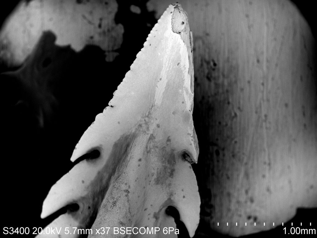
The tip of the barb is the sharpest part of the structure and allows the animal to impale predators.

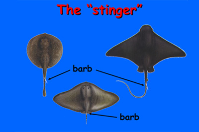
Although some ray species do not have stingers there is a large portion of the group that have them. These structures are not used to hunt for prey but rather as a way to defend themselves from predators. Occasionally people have interactions where they are stabbed by them and it can be a serious situation.


The manta ray (left) is a species that is well known and is absent of a barb. The barb is commonly located at the base or close to the base of the tail (right)

Back in April of 2015 I had an interaction with a bat ray where I was stung in the arm. The initial pain was brief and my adrenaline began to kick in. I asked my friend to take the barb out immediately and thinking about it I may have wanted to keep it in. When thinking about the barb it’s in the shape of a Christmas tree almost. The barb has a sharp point in the top and it’s followed by a series of serrations facing the opposite side of the initial point and taking it out could do more damage than leaving it in, but I was in survival mode and I wanted it out of my body.


This is an image showing the anatomy barbs come in. The darker sections labeled with the number 1 is where the venom glands are located. The number 2

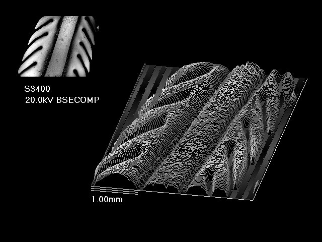
For this post I wanted to look at a barb under the SEM from the same species that stung me and get an appreciation for this defense mechanism. As seen in the second and third picture there are variations in shape and size and it helps with species identification if that is wall that you have to work with. In my ichthyology class we dissected bat rays, where I collected one of the barbs. I also used the Oblique analysis tool to get a three dimensional image of the barb. Can you tell where the venom gland would be located on the barb in relation to picture 3?


These are the images of the barb that specifically focuses on the serrations. The serrations on this structure help the ray increase its capacity of damage if the barb is pulled out.


Stepping into the ring(s)

On the first day of our Scanning Electron Microscopy class our instructor asked if we would like to prepare a sample for the following week. I initially didn’t have a clue on what I wanted to use because I had not prepared any sample of my own to go through the process to be prepped for the SEM. While I was waiting and thinking about what I wanted to use, I realized that I had a pair of otoliths in my backpack. These otoliths were collected from a rock fish during a series NOAA surveys conducted off the West Coast of the U.S. I had them as mementos of the trip I was on and forgot about them for a couple of months and I thought this would be a great opportunity to look at one of them under a high powered microscope.

Otoliths are boney structures located behind the brain of fish and come in different shapes and sizes. Otoliths are not found in cartilaginous fish like sharks, skates, rays and chimaeras and are predominantly in bony fishes. These hard structures are made of calcium carbonate and aid fish in detecting balancing, and directional movement. Scientist can use these structures to identify species, calculate growth rates, and determine the age of the fish. This is made possible because during the lifespan of these fish proteins and calcium carbonate layer on top of each other annually.

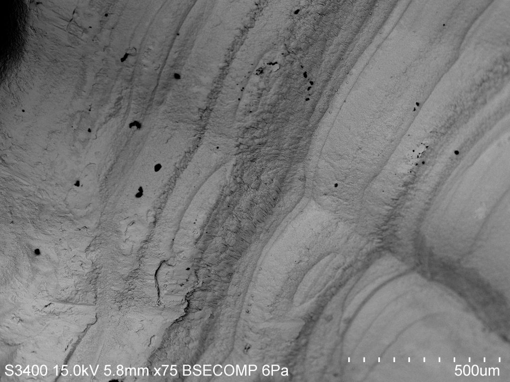
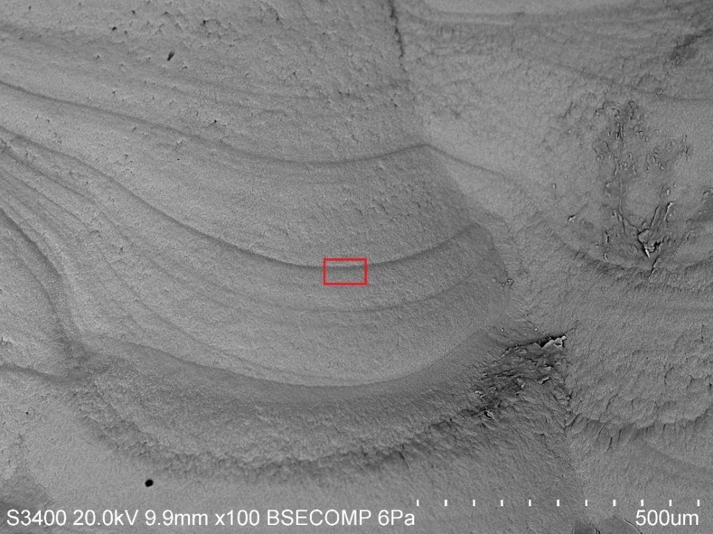
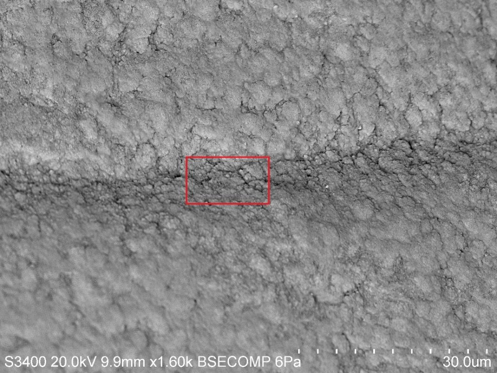
(Picture 1) A comparison in size of an otolith and a penny. Although it’s coated in a thin layer of gold you can see the distinct bands that surround the otolith. These distinct dark “rings” are made of calcium carbonate and proteins, are commonly used to age fishes. This technique is analogous to counting the rings of a tree to determine the age but I was curious to see if there was physical difference between the bands or if it is just a difference in color. When we actually looked at the otolith it almost looked like the surface of a planet. Some areas of the otolith looked like canyons and it was interesting seeing how distinct the bands looked compared to the lighter areas of the otolith. When we looked even closer it seemed that there was a courser texture in the bands than areas without it and the structures were smaller. I thought it was an interesting thing to see not only that there is a color difference but a physical difference in a micro scale.

(Picture 2) A first glance at the surface of the otolith where distinct banding paterns are easily seen. The box is a lead-in to pictures 3&4 where we get a deeper understandig of the physical aspects of the bands.

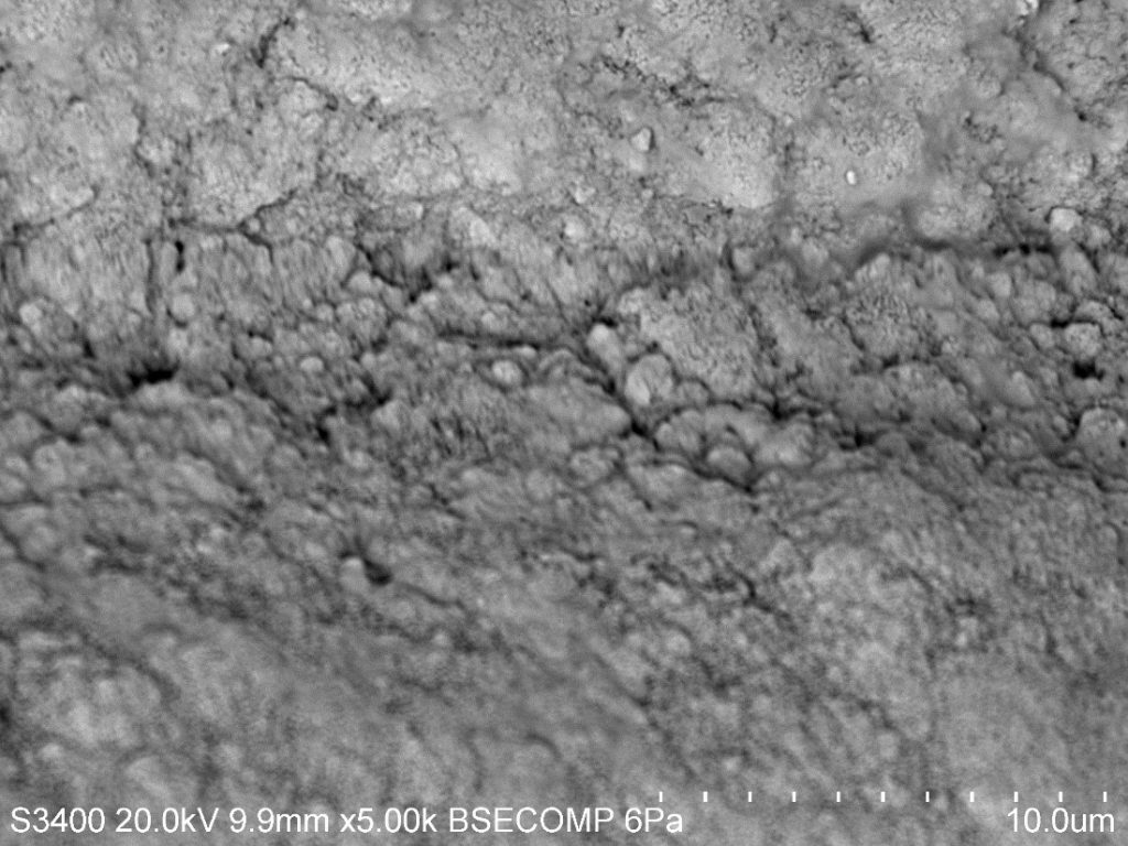
(Picture 3) The physical differences between the darker band and the lighter bands. The red box leads into the physical aspect of the smaller calcium carbonate structures in Picture 4.

(Picture 4) The structures in the band almost mirror the structures on the light side but are smaller and more compact.

Sea-Urchin’ for Detail at a Micro Level

Echinoderms are an interesting bunch of critters, encompassing intertidal favorites such as the star fish, the sea cucumber, and the sea urchin. The roots of the word "Echinoderm" stem from the Green word "ekhinos," meaning porcupine or hedgehog, and the root -derm, meaning skin. In my opinion, sea urchins represent the epitome of a spiky-skinned animal, and in light of that fact I thought I'd look at a few spines under the SEM.

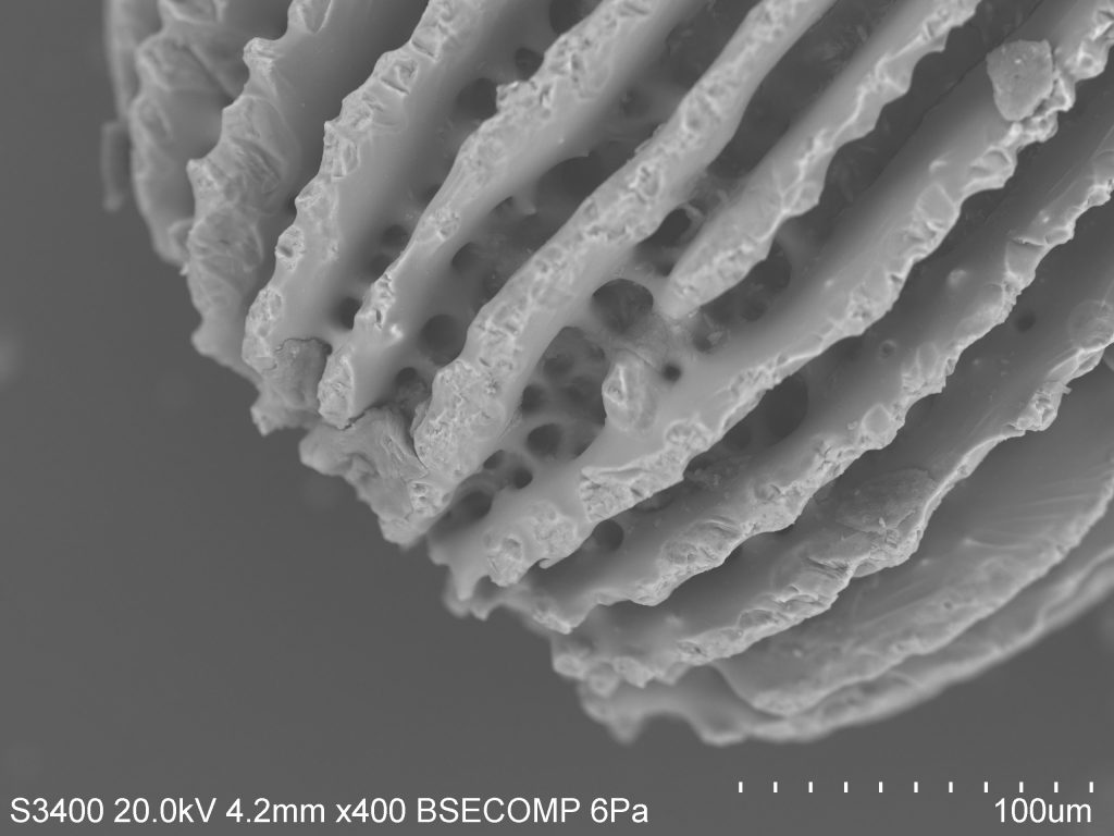
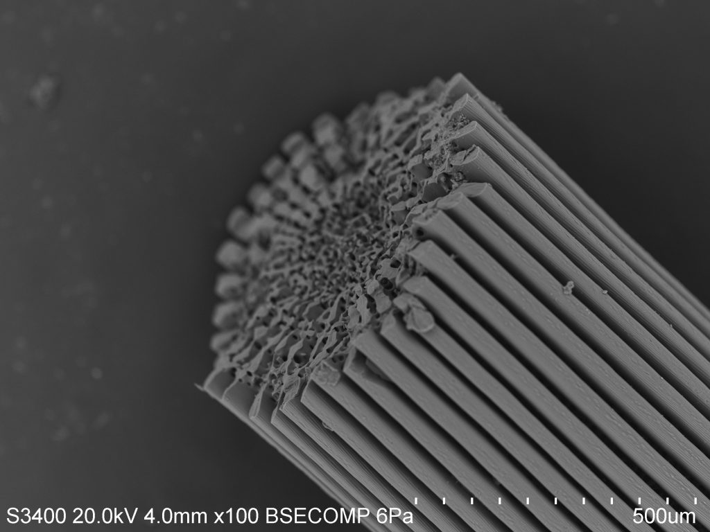
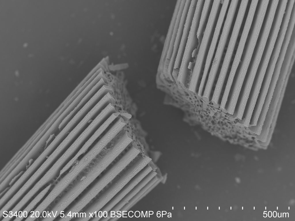
As you can see, the spines are ridged and porous. Sea urchins do not have a closed circulatory system and instead rely on an open water vascular system to help with the uptake of nutrients, the flushing out of wastes, locomotion, and respiration. As such, much of their body plan is open to the environment. In this case, it is actually covered by a thin layer of epidermis (the outer layer of cells), making the spines themselves a feature of the interior endoskeleton. You can think of this as similar to the skin that covers our bones, although spines and bones serve very different purposes.


The name for this internal structural organization is the stereom. The stereom is a feature that all echinoderms share, and is useful in identifying lineages in fossilized samples. Spines mainly show two distinct morphological configurations: the base, made of a meshwork stereom, and the shaft, which has several longitudinal septa and a central core of meshwork stereom. This can be seen distinctly in the photo. The stereom is sponge-like and varies in composition by species. In a sea urchin, the stereom may be 50% living cells such as connective tissue cells and phagocytes involved in nonspecific defense. The rest will be composed of a matrix of calcite sclerocytes which direct mineral production and repair.

Over several days, sea urchins are actually able to regrow spines in places where damage has been inflicted, or to grow entirely new spines. The urchins begin growing micro-spines in a conical shape by precipitating calcite from the surrounding seawater. As these small spines thicken, lateral growth takes place, allowing neighboring micro-spines to join in a horizontal "bridge." The formation of bridges results in the mesh of cells spanning from the base of the spines to the tip.


All this terminology is only the tip of the iceberg (tip of the spine?), and I'm keen to look further. The test of the sea urchin, which is the "hard shell" under the spines and feet and skin, is full of microscopic pores through which several accessory structures pass. The geometry is quite interesting from a macro-perspective, as all echinoderms share pentaradial symmetry, meaning their bodies are grouped loosely into five identical parts. In urchins, these manifest as alternating ambulacral (related to the feet) and inter-ambulacral (between the feet) regions. I imagine the micro-analysis of differences between these regions would be interesting as well.