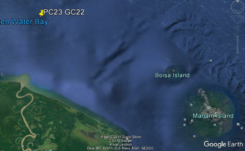
Since we have covered fundamental information regarding microfossil composition and structure, now we can focus on individual core sites and general geology of the New Guinea region. The science crew aboard the Roger Revelle (RR1313) collected a total of 54 cores near the Papua New Guinea margin. Sites PC23 and GC22 are the southeastern most sites, situated east of the Sepik Delta and west of the Manam Volcano. We will be focusing on piston core site 23 (PC23) collected at a water depth of 712 m and a subsurface depth of 431-432 cm.
Tectonically, the New Guinea region is complex due the formation of a subduction zone by rapid (geologically speaking) motion of the sea-floor. Subduction, indicated on the map as black lines with the teeth, is a geological process that occurs when two plate boundaries collide. As the two plates collide the denser slab sinks into the mantle and beneath the lighter slab, where temperature and pressure progressively increase with depth. Under these conditions trapped fluids such as seawater generates magma, which rises into the upper slab, and forms a volcano. In this case, the Indo-Australian Plate is moving northeastward but crashes into the Pacific Plate that is moving west with a slight north component. This vertical and horizontal displacement is geometrically classified as oblique-slip faulting, resulting in the formation of a volcanic chain.
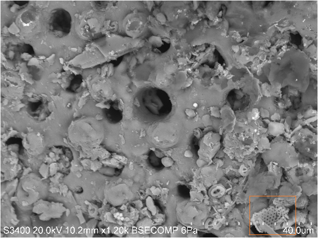
Sediment at PC23 is particularly exciting because it is biologically and lithologically diverse. The image on the right displays several coccolithophores on top of flaky clay-sized particles. The image on the left features several coccolithophores that have situated on top of a “hole bearing” organism, known as foraminifera. A smaller foram fragment (orange) is visible in the lower right. Looking at sediments on a SEM is especially useful for end-member microanalysis. Mineral end-member identification is used to distinguish between biogenic, terrigenous, and hydrothermal sediments, all of which may be altered when water masses mix and interact with rock.
An EDX detector paired with the SEM is able to generate data in the form of a spectra that displays known elements in the point scan by focusing a high-energy beam of particles into the sample. The output spectroscopy peaks for Spectrum 2 above indicates that calcium is present (peak intensity does not correspond with weight %), verifying that the microfossil analyzed is a coccolithophore.
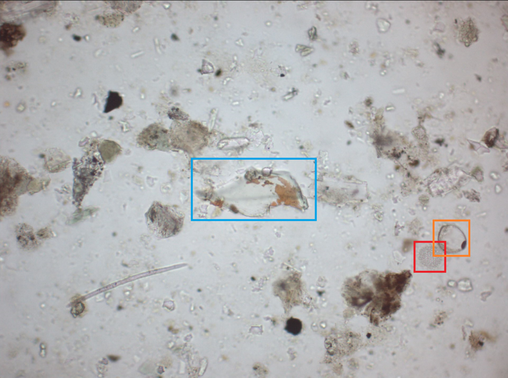
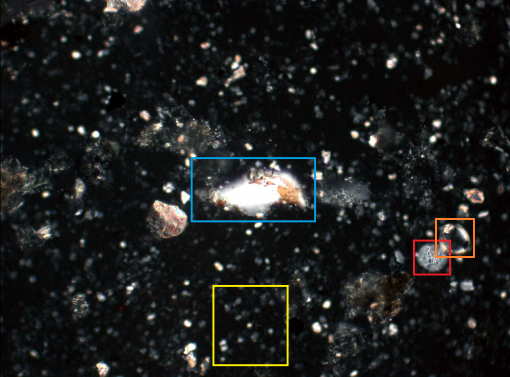
In addition, scientists can analyze sediments underneath a petrographic microscope by preparing smear slides. A smear slide is a thin layer of unconsolidated sediment spread onto a glass slide. Petrographic analysis of a smear slide gives quantitative and limited compositional information with the ability to view the sample in plane-polarized light (PPL, top) and cross-polarized light (XPL, bottom). Under PPL, the sediment from PC23 appears to be clay-rich and contain minimal biogenics. However, under XPL we can see that the sediment is abundant in biogenics relative to the other particles. Coccolithophores (cluster in yellow) are calcite bearing organisms that are only visible in XPL. The dinoflagellate (red) and foraminifera (orange) are visible under both light conditions. The centered shard of vitric material (blue) is an indicator of hydrothermal activity and suggests volcanism. The combined use of a SEM and petrographic microscope make for strong tools to reconstruct ancient oceans.


