Hydroid

Today, we're going to look at a member of the Cniderian phylum, named after the Greek word for "nettle" in reference to their special stinging cells, the nematocyst. You probably think of jelly fish when you think of stinging sea creatures, but Cniderians consist of four subgroups: the Scyphozoa (jellies), the Cubozoa (box jellies), the Anthozoa (sea anemones), and Hydrozoa (hydriods).

Most hydrozoans are fairly inconspicuous. You probably know them as the fuzzy masses you see growing on pilings and boat-bottoms. While most are fairly harmless, there are some capable of delivering a fairly painful sting. For this discussion we're going to be focusing on colonial hydroids. In these communal groupings, individual polyps are connected and share resources through a hydrocaulus. Colonial hydriods have specialized individuals, or "zooids," that exist to fulfill a specific need in the colony. For example, the most common type of zooid is a gastrozooid, which provides the colony with food. Gonozooids are used in reproduction, while in some species nematocyst-filled cells called cnidocytes aid in defense.

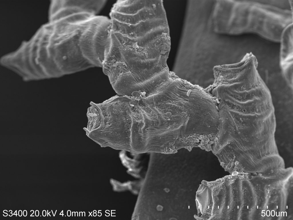
These SEM photos below show the chitonous exoskelton, or perisarc, of the colonial hydroid Obelia. This species is well-studied by scientists and is commonly used in zoology classes to describe the basic hydroid body plan and life cycle. The main body of the colony is composed of living tissue known as the coenosarc, which is covered by the perisarc. Colonies can grow in a variety of patterns, which are often used for identification purposes. Obelia, for example, demonstrates the branching pattern some hydroids adopt, while others grow vertically off of a common stolon.


Both body forms indicative of the greater Cniderian grouping, the medusa and the polyp, are present in this species. Medusae are released from the gonozooids, producing free-swimming medusae velum with gonads, a mouth, and tentacles. The medusae reproduce sexually, releasing sperm and eggs that fertilize to form a zygote, which later morphs into a blastula, then a ciliated swimming larva called a planula. The planulae live as free-swimming members of the planktonic community before eventually attaching themselves to a solid surface, where they begin their reproductive phase of life. Once attached to a substrate, a planula quickly develops into one feeding polyp. As the polyp grows, it begins developing branches of other feeding individuals, thus forming a new generation of polyps by asexual budding.


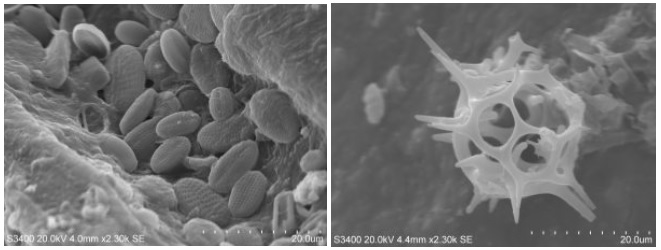
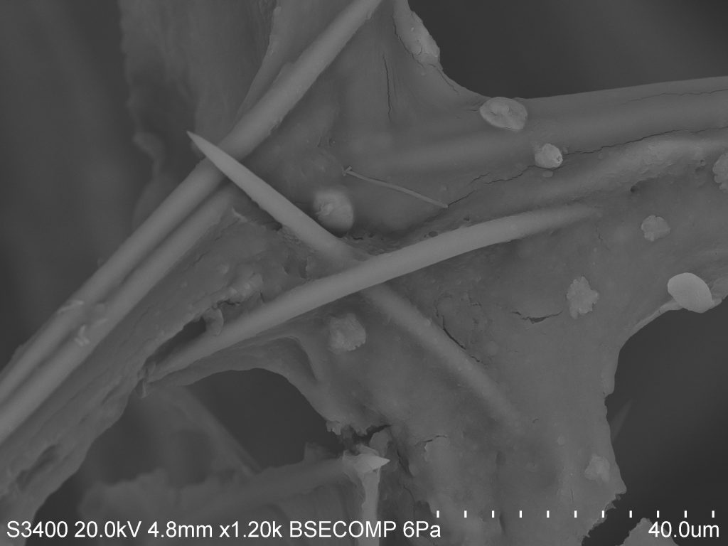
If you read Elizabeth's recent post regarding crustose coralline algae, you saw that it's very common to find unexpected organisms associated with your target species. As I was imaging this hydroid sample, I came across a few stow-aways that were too interesting not to include. On the left is a cluster of diatoms, marine phytoplankton responsible for much of the primary production in our oceans. They are often used in analyzing sediments, as discussed in some of Jennifer's posts in this atlas. On the right is an unidentified planktonic organism. Even when it's not possible to ID something on site, we can use these images to make inferences regarding their life history. For example, spines such as these are thought to function in decreasing the rate of sinkage for these organisms. At such small sizes (not the scale), water represents a very viscous medium, and increasing surface area is one way to create drag and stay afloat.


Sea-Urchin’ for Detail at a Micro Level

Echinoderms are an interesting bunch of critters, encompassing intertidal favorites such as the star fish, the sea cucumber, and the sea urchin. The roots of the word "Echinoderm" stem from the Green word "ekhinos," meaning porcupine or hedgehog, and the root -derm, meaning skin. In my opinion, sea urchins represent the epitome of a spiky-skinned animal, and in light of that fact I thought I'd look at a few spines under the SEM.

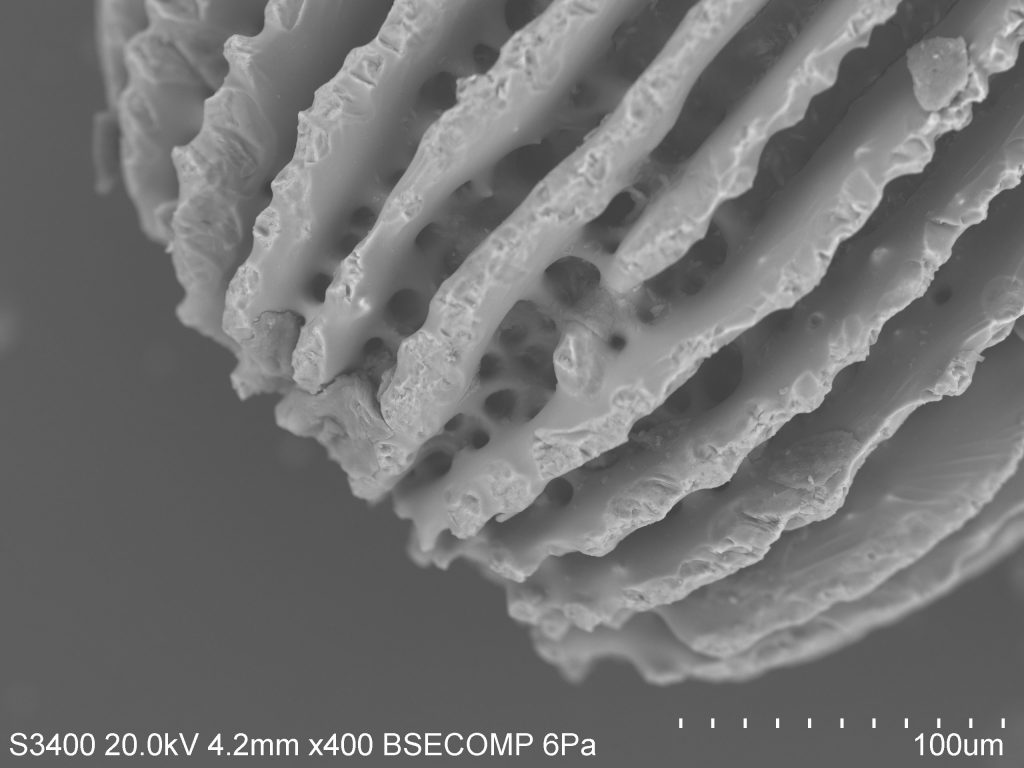
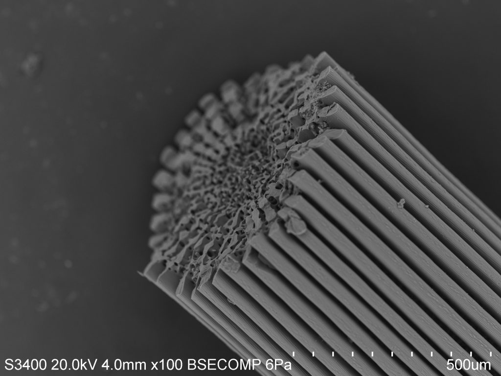
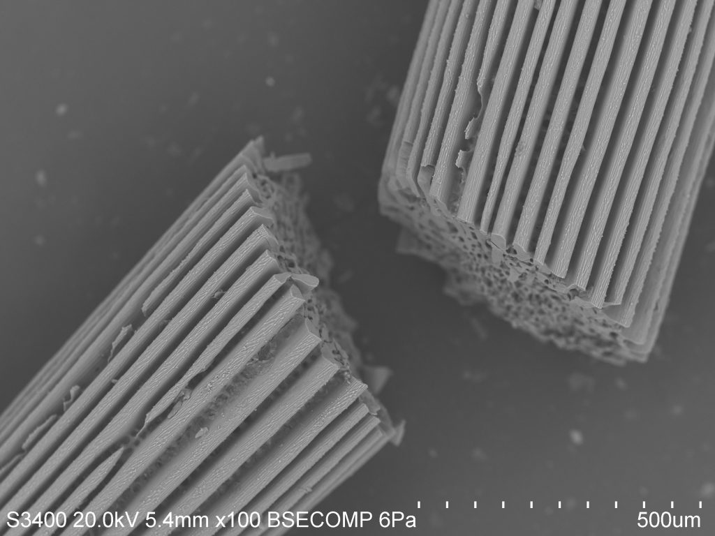
As you can see, the spines are ridged and porous. Sea urchins do not have a closed circulatory system and instead rely on an open water vascular system to help with the uptake of nutrients, the flushing out of wastes, locomotion, and respiration. As such, much of their body plan is open to the environment. In this case, it is actually covered by a thin layer of epidermis (the outer layer of cells), making the spines themselves a feature of the interior endoskeleton. You can think of this as similar to the skin that covers our bones, although spines and bones serve very different purposes.


The name for this internal structural organization is the stereom. The stereom is a feature that all echinoderms share, and is useful in identifying lineages in fossilized samples. Spines mainly show two distinct morphological configurations: the base, made of a meshwork stereom, and the shaft, which has several longitudinal septa and a central core of meshwork stereom. This can be seen distinctly in the photo. The stereom is sponge-like and varies in composition by species. In a sea urchin, the stereom may be 50% living cells such as connective tissue cells and phagocytes involved in nonspecific defense. The rest will be composed of a matrix of calcite sclerocytes which direct mineral production and repair.

Over several days, sea urchins are actually able to regrow spines in places where damage has been inflicted, or to grow entirely new spines. The urchins begin growing micro-spines in a conical shape by precipitating calcite from the surrounding seawater. As these small spines thicken, lateral growth takes place, allowing neighboring micro-spines to join in a horizontal "bridge." The formation of bridges results in the mesh of cells spanning from the base of the spines to the tip.


All this terminology is only the tip of the iceberg (tip of the spine?), and I'm keen to look further. The test of the sea urchin, which is the "hard shell" under the spines and feet and skin, is full of microscopic pores through which several accessory structures pass. The geometry is quite interesting from a macro-perspective, as all echinoderms share pentaradial symmetry, meaning their bodies are grouped loosely into five identical parts. In urchins, these manifest as alternating ambulacral (related to the feet) and inter-ambulacral (between the feet) regions. I imagine the micro-analysis of differences between these regions would be interesting as well.


Sponges and Spicules

We've dabbled a bit with invertebrates thus far, talking about house flies and various planktonic organisms, but now we're going to work our way into some more "visible" representatives, even if they often get overlooked. That group, the phylum Porifera, represents the 8,755 valid species of sponge, most all of which are marine. While some sponges are very colorful (such as the Caribbean Blue Sponge) or very large (Giant Barrel Sponge), most sponges are small or cryptic and require very fine-scale analysis for proper species identification. Seems like a perfect application for SEM technology! Continue reading below for an introduction to spicules (the "bricks" of sponge architecture) and how they can be used to identify individual species.

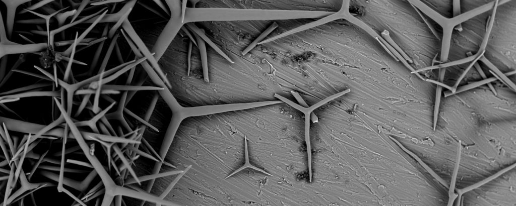
Some terminology, to begin, because sponge taxonomy is a whole other language in itself even by science standards. The exoskeleton of sponges (so, the parts that you see) are composed of a mixture of  spongin and/or spicules. Spongin is a modified type of collagen protein, and forms the "fibers" or "mortar" that hold spicules together.  Generally, species are identified based on the presence or absence of spongin in a sample. Spicules are the structural components of a sponge, or the "bricks," and the shapes, sizes, and composition are unique for each species. Together, you can look at these features under a microscope to make a positive identification.


Spicules are composed of either Calcium or Silica. Looking at composition is another way to narrow down possible sponge groupings. The "brightness" of the sample under the SEM is one way to guess at content. Calcium has a higher atomic number than Silica and is a better conductor, so it appears brighter. However, we recently learned how to use the Energy-Dispersive X-ray (EDX) function of our SEM. EDX detects and measures X-rays generated by a sample, the specifics of which are determined by elemental composition. With this tool, we were able to determine the elemental makeup of several geographically diverse samples.

Left: a siliceous deep-sea sponge whose spicules form discrete hexagonal patterns, the bottom of which you can see. Right: a calcareous semi-tropical sponge with a loosely-formed spicule matrix.

This is a very basic description of sponge identification, but there are loads of resources available on the internet, and spicules themselves can be quite beautiful. For more information, please feel free to visit:

  1. Shape of Life for short videos about sponges and their spicules
  2. The World Porifera Database for recent publications, photos, and information
  3. Southern California Association of Marine Invertebrate Taxonomists for the aforemetioned "dictionary" of spicule morphological terms

Foraminifera and Coccolithophores

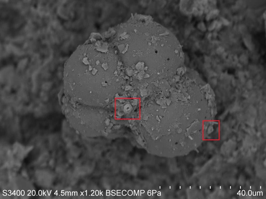
Chances are you'll be seeing a fair bit of discussion regarding plankton as we go forward, because (1) they show up in many of our samples, and (2) there's a lot we can learn by studying them! SEM is especially useful for the study of plankton, as their small size makes traditional compound microscopes somewhat ineffective. This photograph, taken during our first run-through with the SEM, shows an image at 1,200X magnification, as compared to the 100X magnification you might hope to get with a standard scope!


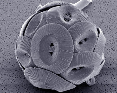
In the above photo we can see examples of both zooplankton (microscopic animals) and phytoplankton (microscopic plants). The first is the obvious, bulbous species in the middle of the image. This is the test, or shell, of a Foraminifera. Forams represent an ancient and speciose group of zooplankton which live mostly in sediment (as is the case here), but also in the water column. Within the red squares you will see a second, smaller phytoplankton species known as a Coccolithophore. The round plates are known individually as placoliths, and bundle together around the algal species to form a coccosphere. I've included the photo below to give you an idea of how they appear when alive in nature. Similarly, coccolithophores inhabit both benthic and open-water habitats.


Plankton species such as these can provide a surprising amount of information not only about themselves, but also about the environment in which they grow.  The tests you see here are made of calcium carbonate (CaCO3) which the organisms "pull" out of the water and use to build their hard parts. As such, isotopic analysis of these tests can tell you about the atmospheric and oceanic conditions at the time of their formation, since the two systems are linked in the carbon cycle. When the organism dies, calcium carbonate sinks to the bottom of the ocean in the form of coccoliths and other tests and become a part of the sediment record, which can be studied later by scientists as part of a sediment core.

There are many other applications in the study of plankton, some of which we will likely cover in future installations. Stay tuned, and remember: It's the little things.

Common Housefly

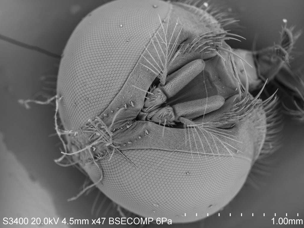
This SEM image of a housefly at 47x magnification highlights the intricacies possessed by even the most common lifeforms. One may be intrigued by the stark contrast between the fine hairs and the thousands of hexagonal structures making up the eyes. Together the eyes create a mosaic-like image, giving flies spectacular peripheral vision. This may seem like a huge visual advantage. However, flies are unable to focus; they can see motion and form, but are unable to clearly see objects. This is why flies are so quick to flee. They see something headed their way, but have no clue what it may be.


The hexagonal shape making up a fly’s vision is not unique in the natural environment. This structure is found in numerous natural features such as honeycombs, columnar basalt, turtle shells, snakeskin, etc. This structure is so commonly found due to its geometry. Triangles, squares, and hexagons are the only three shapes with equal sides and angles that fill a flat plane. Of these, hexagons require the least total length of wall, making it the more efficient shape.


Marine Bacteria on Top of a Diatom Chain

Bacteria = rod shaped cells approximately 1-2µm (smaller than blood cells).

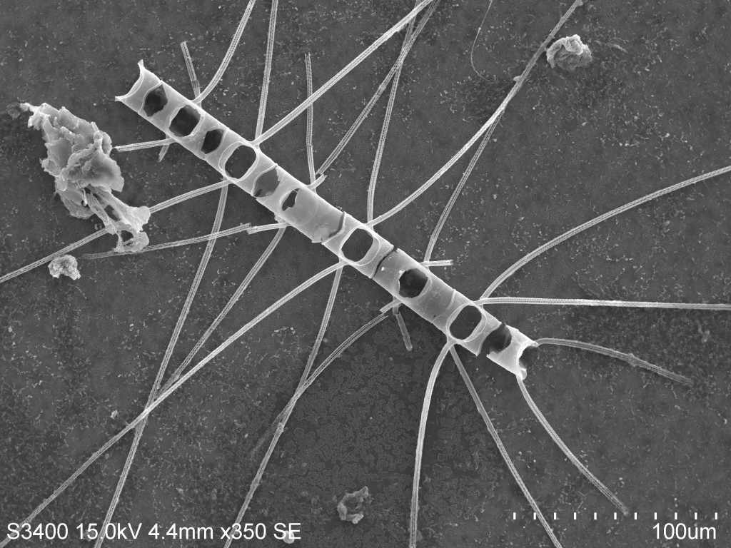
Bacteria sit atop a diatom chain (Chaetoceros unknown sp.) absorbing dissolved organic carbon, enjoying the 28C surface water temperature, and undergo binary fission whenever they want. Unbeknownst to them, the bacteria are scooped up in the middle of the Atlantic by Jules Kuo and Jasmine Ruvalcaba (recent graduates of MLML) using a zooplankton net that captures organisms >50 µm.  The bacteria are around ~1-2 µm but they were able to be captured as they have glommed onto this chain forming diatom.  Though the individual diatoms themselves are 10 µm in diameter, they can form chains containing more than 12 individuals at a time (~120 µm).