The birds and the bees according to coralline crust

Have you ever wondered how algae reproduce? Algal species do not reproduce like plants specifically by using pollen and most do not have special friends like bees to help them reproduce with other individuals. The cool thing about algae is that each kind of algae and sometimes even individual species experience the "birds and the bees" in a different way. For instance, there are some species of algae that belong in the green group that have positive and negative phototaxis gametes. This means that when the gametes (male or female) need to find a mate, they are attracted to the light and come to the surface (positive phototaxis), where they meet all the other singles in the area. Kind of like going out to a bar. Once they have found their mate, they sink to the bottom (due to negative phototaxis) to settle together on a rock and make a new individual. Kelps, belonging to the brown algal group, have special pheromones and flagella that are used in the reproductive process. Female gametophytes or eggs have different kinds of pheromones like animals do that attract the sperm to the egg. Essentially, kelps make the boys do all the work! The males have flagella, or swimming appendages, that help them move towards the female once they recognize the pheromone. Once the female gametophyte is fertilized the female is taken over and the new individual actually grows from that female egg. Red algal species are also very different in how they reproduce.

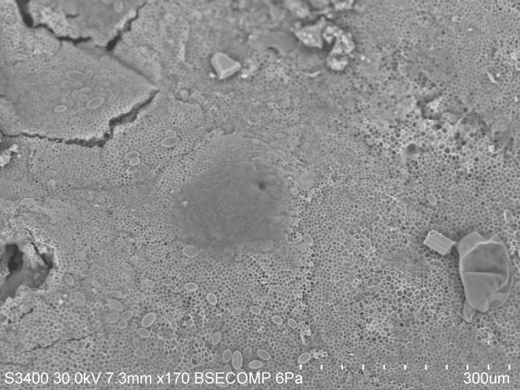
Specifically, coralline algae, the crustose form, has to get very creative in its reproductive cycle. When the coralline crust is ready to reproduce it creates little cenceptacles or cases along the top of the crust. It then fills the conceptacle with reproductive material or tetrasporangia. When they are ready to release this material, the conceptacle bursts through the top layer of the crust and the reproductive material, or tetraspores are cast into the water column to find a mate and settle on a nearby rock before something comes along and eats it! In the picture to the right you can see this conceptacle in the crust. It has yet to fully burst.


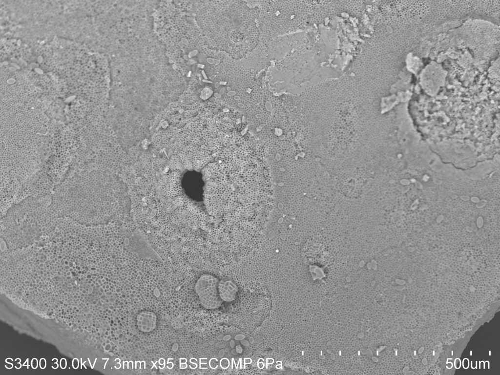
However, the picture on above is on the same piece of crust just in a different area and you can definitely see where the conceptacle used to be. If you were to look at this crust with the naked eye you would not have been able to see any of this. But thanks to the SEM, a human can actually see the detail of an individual conceptacle. To a phycologist, this is SUPER cool! Hopefully you all find it slightly fascinating too!

The inside scoop on Giant Kelp

If you live by the ocean, especially on the California coast, then you are probably familiar with Giant Kelp. This is the species of kelp that creates large brown forests underwater and lovely fly infested lumps of smelly nastiness on the beaches. Giant Kelp, known to phycologists as Macrocystis pyrifera, is one of the most common seaweeds people associate with the ocean. This is because this particular species is found year round. Unlike other seaweeds that tend to have seasonal debuts, Giant Kelp has a special mechanism for growth and reproduces year round, making it resistant to seasonal changes in its environment.

Giant Kelp is a type of algae, not a type of plant like what you would think of on land. Land plants grow using xylem and phloem, two mechanisms responsible for storing water and transferring nutrients throughout the structure, respectively. Algal species are not related to land plants and therefore do not have these same mechanisms for growth. Instead, in algae like kelps, they have what is called trumpet hyphae, which look like a bunch of trumpets that move nutrients throughout the entire thallus. This mechanism is extremely successful. In fact, the Giant Kelp species is so successful at transferring its nutrients that it can, in the best environmental conditions, grow up to a foot a day! This rapid growth helps keep Giant Kelp around during any season, creating the forests that we all know and love.

Giant Kelp is also resistant to seasonality due to its reproductive methods. This particular species has what we call sporophylls which are specialized blades specifically grown to reproduce. Sporophylls are full of reproductive spores and they reside at the bottom of the frond just above the holdfast. It is most obvious that they are ready to reproduce when the blades turn a milky white. For this particular segment I wanted to use the Scanning Electron Microscope to look at the cross section of sporophyll blade. I cut a small square out of the milky sporophyll blade I had collected and stuck it on a stub.
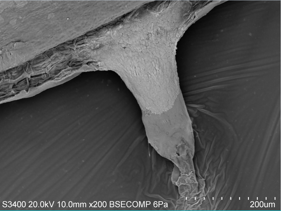
You can see in the picture to the left that you can view the top and bottom of the blade but the focus is on the inside. You can see the honeycomb structure inside the blade. When I scanned over to this part of the cross section I found this odd structure oozing out of my cross section. After chatting with a few of my lab mates we have decided that the sporophyll was oozing out some sugar mucilage. This means that this particular blade was full of good nutrients!


The more I work with algae the more I realize how complicated it is to work with tissue on the SEM instead of hard organic material like some of my class mates. I find it as a fun challenge to continue to understand the microscopic structures of algae!

Coralline Algae…. the unsung hero

Many I am sure have seen the beautiful pink and purple hues that dance in the tide pools during low tide. Some may have also seen pink or purple looking rocks in the sand along Monterey Bay's coast.

These beautiful colors you see are not a kind of coral or a type of rock, contrary to common belief. Their name can be misleading but corallines, crustose and articulated, or upright and branched, are actually types of red algae found in many habitats around the world. They get their name because of the calcium carbonate within their cell structures on their thallus that creates a hard outer form of protection from the environment and hungry grazers. An articulated coralline can be found in the low intertidal and the subtidal. Its hard structure provides protection from breakage when living in areas like the low intertidal where wave energy can be high. Because the structure is not completely rigid, there is just enough flexibility for it to sway back and forth with the tides. The calcium carbonate within their thallus also provides protection from grazing invertebrates. Instead of having a fleshy outer structure, the coralline has a hard rough structure that doesn't seem as appetizing. However, there are a few choice grazers who actually prefer coralline, especially in its crustose form, like chitins that have a rough tongue called a radula that is used to scrape food off of rocks. 

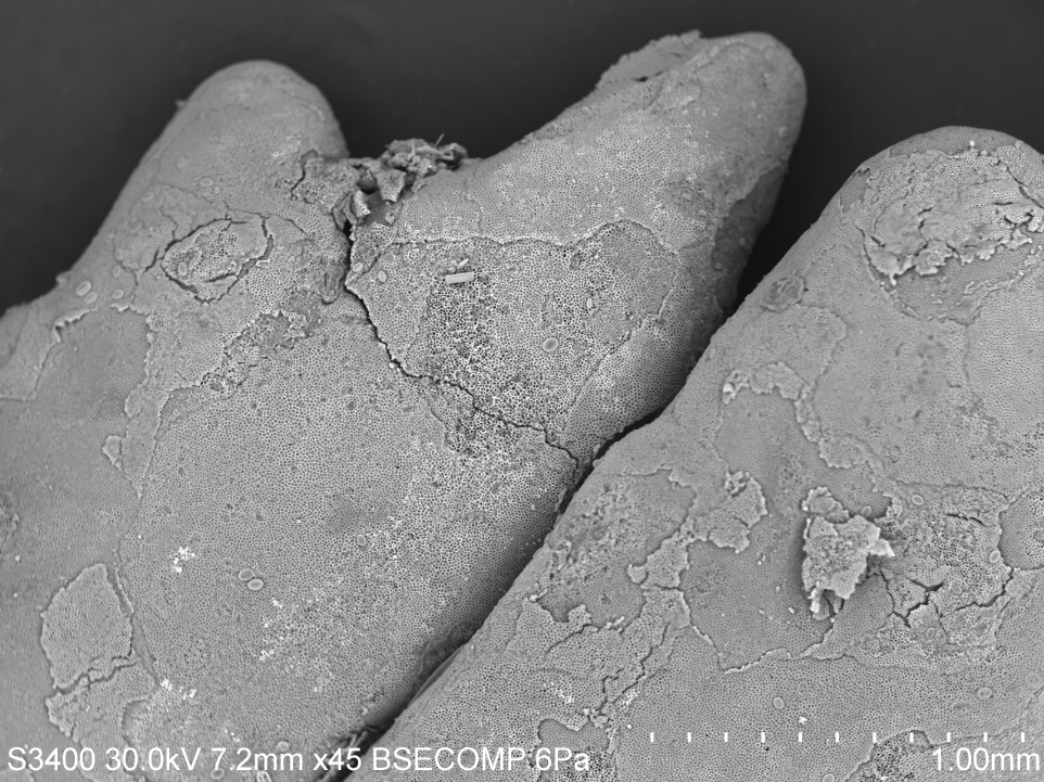
If you look at this SEM photograph to above you can see the calcium carbonate structure well. What you are seeing are individual layers of cells on the outer most crustose shell shedding off. Some species of coralline algae do this to anti-foul or get rid of any epiphytes that may be eating away and hindering the health of the individual. This was first seen using scanning electron microscopy methods and as research continues to evolve, more studies are finding that this is mechanism is quite common especially in crustose species of coralline.

If you look even closer, like at the SEM picture below, you can see the honeycomb structure of the cells. These cells have that hard structure made of calcium carbonate, also known as limestone. What is truly fascinating about coralline algae, espeically the crustose form, is that it is actually an unsung hero for the coral reefs in tropical environments. When the crustose coralline algae settles and starts to grow, it creates a glue or cement that ultimately keeps coral reef beds together. Because of its limestone cellular structure, coralline has been shown to positively influence coral reefs worldwide. If you want to learn more, follow this link.

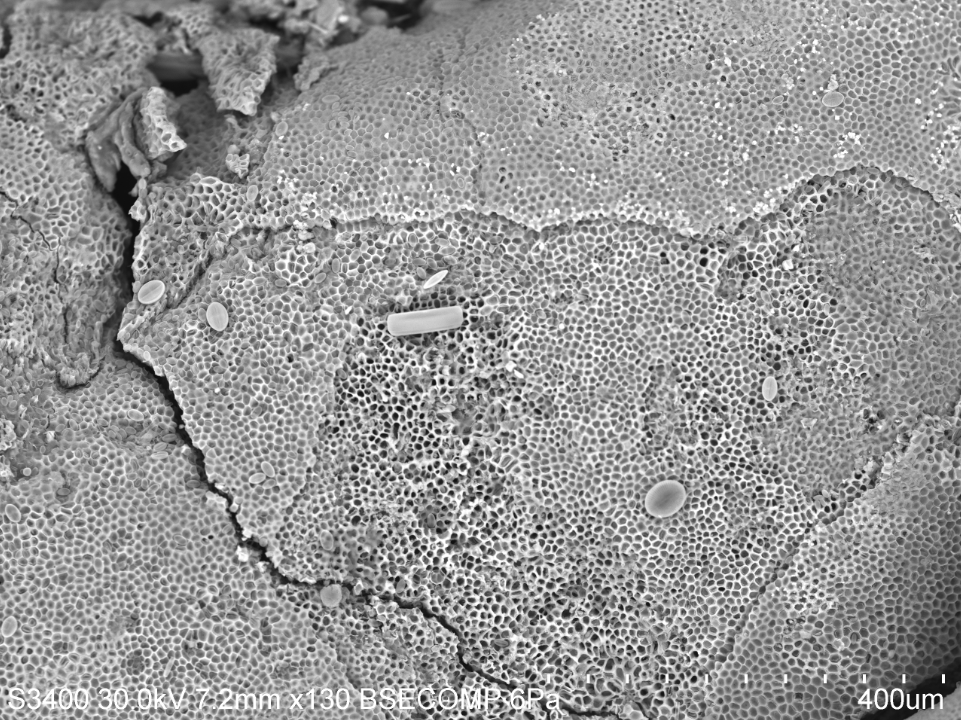
I purposely chose to not wash off the coralline very well before creating a stub so that I could see what kinds of things may have been living on my articulated coralline specimen.
In this picture to the right you can see there are a few geometric shapes on top of the structure. These are all different kinds of diatoms that were either just in the water that was brought in with the coralline or they were living on top of the actual structure. The most exciting thing about using the scanning electron microscope is finding what other microscopic things may be living with your species of interest.