If you live by the ocean, especially on the California coast, then you are probably familiar with Giant Kelp. This is the species of kelp that creates large brown forests underwater and lovely fly infested lumps of smelly nastiness on the beaches. Giant Kelp, known to phycologists as Macrocystis pyrifera, is one of the most common seaweeds people associate with the ocean. This is because this particular species is found year round. Unlike other seaweeds that tend to have seasonal debuts, Giant Kelp has a special mechanism for growth and reproduces year round, making it resistant to seasonal changes in its environment.
Giant Kelp is a type of algae, not a type of plant like what you would think of on land. Land plants grow using xylem and phloem, two mechanisms responsible for storing water and transferring nutrients throughout the structure, respectively. Algal species are not related to land plants and therefore do not have these same mechanisms for growth. Instead, in algae like kelps, they have what is called trumpet hyphae, which look like a bunch of trumpets that move nutrients throughout the entire thallus. This mechanism is extremely successful. In fact, the Giant Kelp species is so successful at transferring its nutrients that it can, in the best environmental conditions, grow up to a foot a day! This rapid growth helps keep Giant Kelp around during any season, creating the forests that we all know and love.
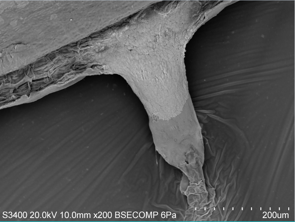
Giant Kelp is also resistant to seasonality due to its reproductive methods. This particular species has what we call sporophylls which are specialized blades specifically grown to reproduce. Sporophylls are full of reproductive spores and they reside at the bottom of the frond just above the holdfast. It is most obvious that they are ready to reproduce when the blades turn a milky white. For this particular segment I wanted to use the Scanning Electron Microscope to look at the cross section of sporophyll blade. I cut a small square out of the milky sporophyll blade I had collected and stuck it on a stub.
You can see in the picture to the left that you can view the top and bottom of the blade but the focus is on the inside. You can see the honeycomb structure inside the blade. When I scanned over to this part of the cross section I found this odd structure oozing out of my cross section. After chatting with a few of my lab mates we have decided that the sporophyll was oozing out some sugar mucilage. This means that this particular blade was full of good nutrients!
The more I work with algae the more I realize how complicated it is to work with tissue on the SEM instead of hard organic material like some of my class mates. I find it as a fun challenge to continue to understand the microscopic structures of algae!


