Today at the microscope: Diatom and Coccolithophore BFFs

Yesterday as I was looking under the microscope at particles collected from the bottom of the ocean, and I came across this beautiful cell(s):
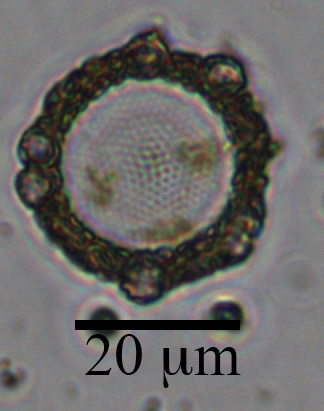
 

This centric diatom is surrounded by a halo of smaller coccolithophore cells.  The circle in the center is the diatom.  You can see the nano-patterns on the silica valve and some yellow/brown-ish chlorophyll inside the cell.  Surrounding the cell are atleast 6 coccolithophores cells (smaller circles) and a ring of their coccoliths (the brown-ish looking mass).  The silica frustules of diatoms and calcium carbonate coccoliths of coccolithophores are relatively heavy biominerals, and may increase carbon export out of the surface ocean by “ballasting” sinking particles.  There is some debate in the literature about which biomineral is more important in exporting carbon.  This diatom-coccolithophore association illustrates my opinion: they are probably both very important.

This sample was collected in a sediment trap 4000 m deep, at the Station M time-series.  I am currently counting phytoplankton within the particles collected over the past 30 years in these sediment traps in collaboration with Ken Smith and Crissy Huffard at MBARI, and thanks to support from  a 2017 New Faculty Award from the California Sea Grant.

Read more from Sea Grant here:
https://caseagrant.ucsd.edu/news/special-focus-awards-support-new-faculty-resilient-coastal-communities
https://caseagrant.ucsd.edu/project/how-does-climate-change-affect-the-export-of-phytoplankton-to-the-seafloor

New microscope, ready to bring out to sea!

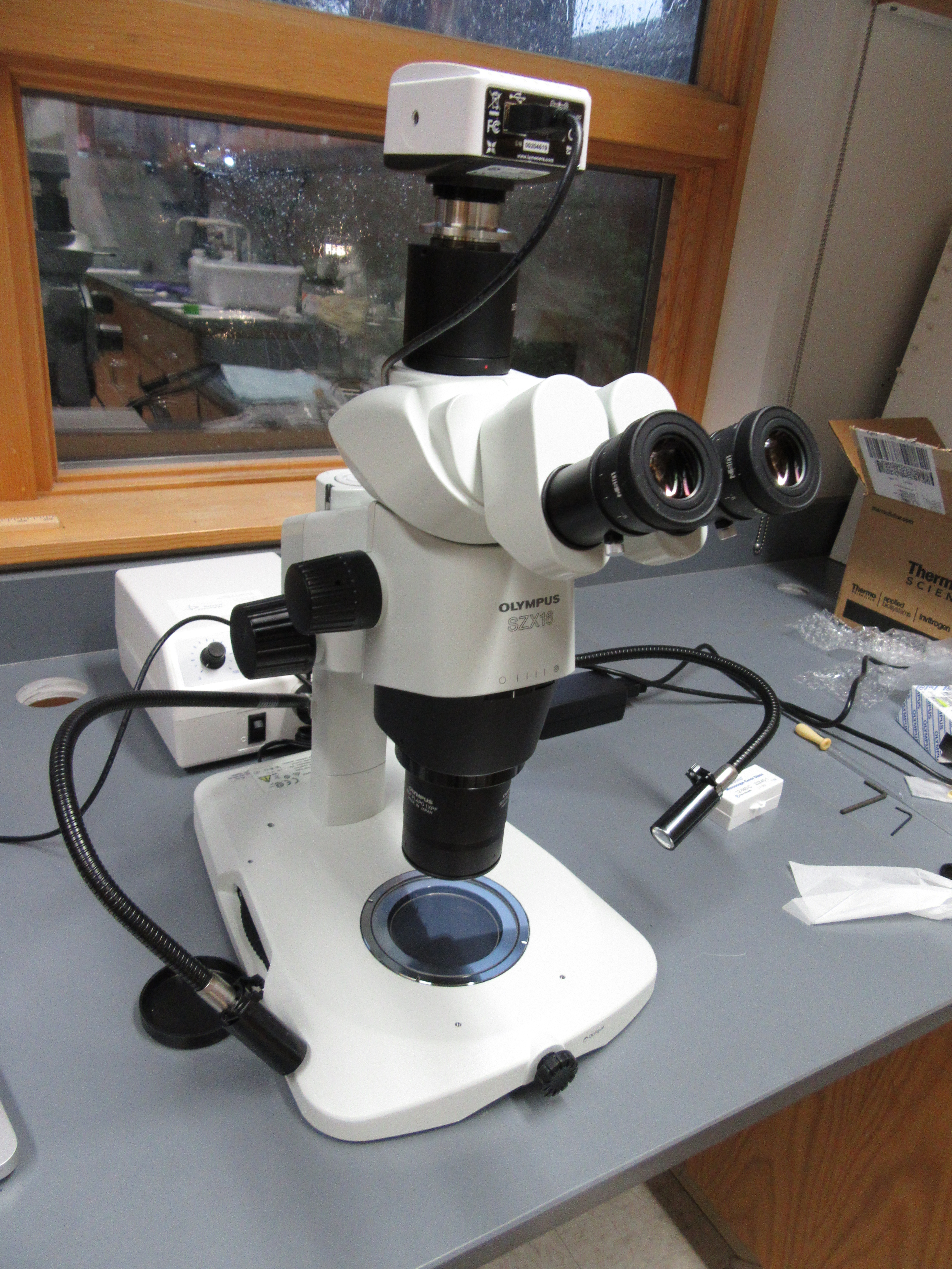
This week I received my lab’s first microscope!  It is an Olympus SZX16 stereo microscope. Stereo MicroscopeThis microscope will be used to image and quantify sinking particles collected in sediment traps.  It also fits easily into a carry-on size bag so that I can safely transport it out to sea.
img_0002


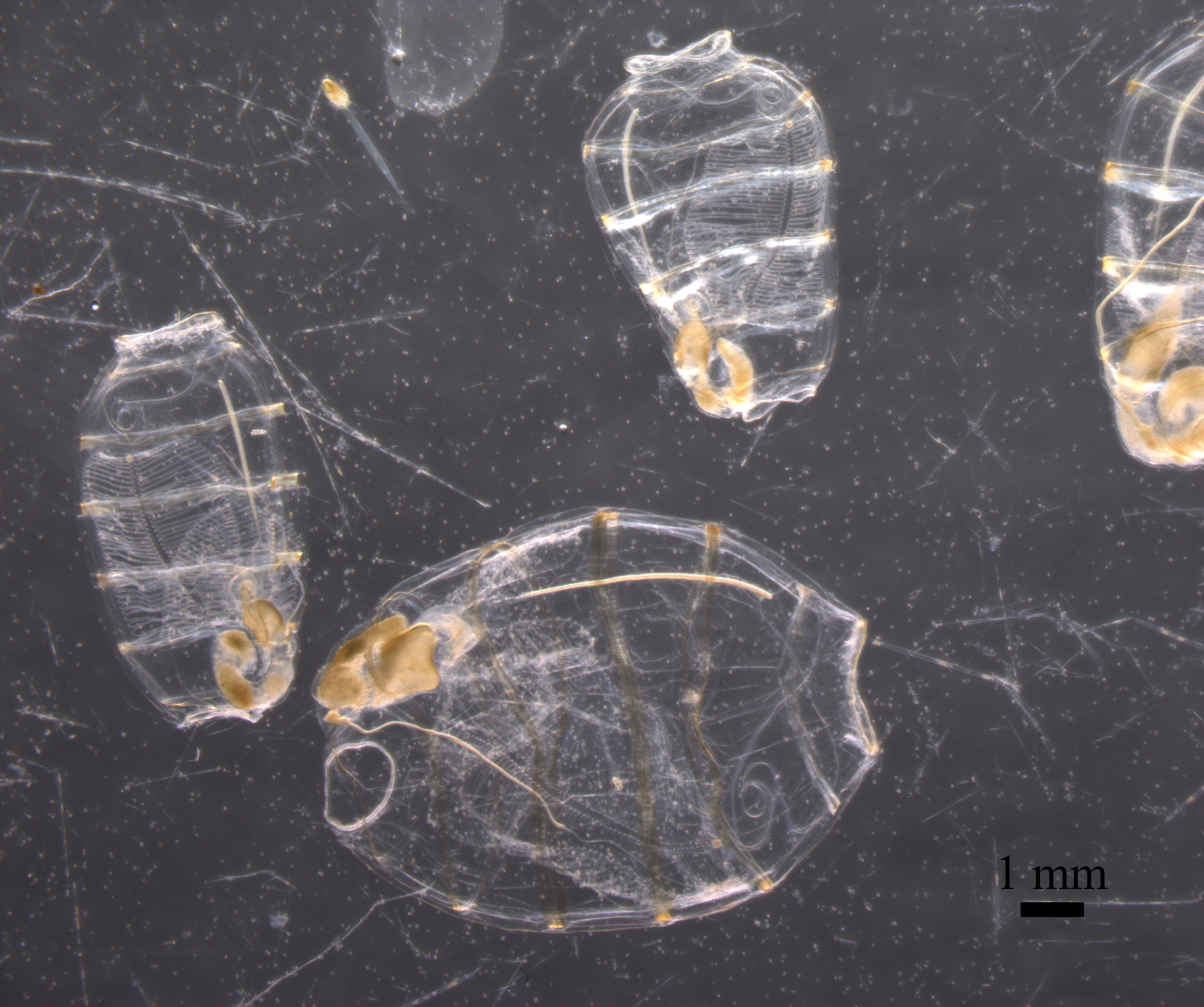
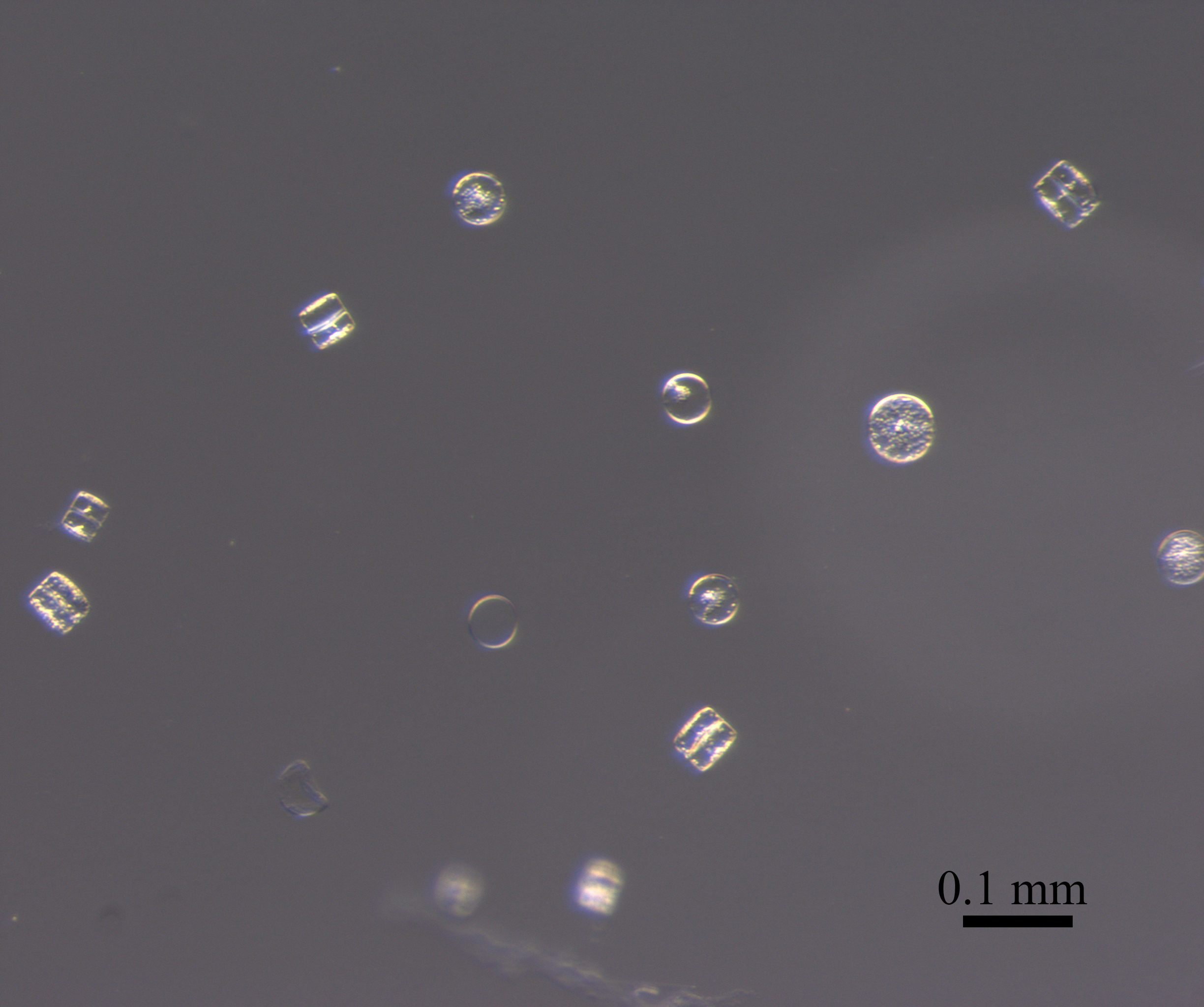
This microscope is coming with me on the upcoming Sea2Space cruise aboard the R/V Falkor (leaving next week!), and is part of a new research project recently funded by NSF.  I received it just in time to test out its imaging capabilities with a preserved plankton net tow I collected last summer in Monterey Bay.  This sample contained a type of gelatinous zooplankton called doliolids and a bloom of diatoms.  I love how we can see both large and small things:

The doliolids are between 0.5 cm and 1 cm long, while the diatoms are about 0.005 cm long (or ~50 micrometers).  The diatoms in this image are called Coscinodiscus.

diatoms doliolids

I can’t wait to image sinking particles next month with my first microscope, and I’m already dreaming about microscope #2 for the lab.

Diatom community composition and aggregation

Posted by CSUMB REU student Melia Paguirigan:

This summer I participated in the California State University Monterey Bay Research Experience Undergraduate program, with Dr. Colleen Durkin as my mentor. Our project investigated the role of diatom community composition and morphology in aggregation. We collected whole seawater samples from Monterey Bay.

Durkin_Paguirigan_Whalercollection
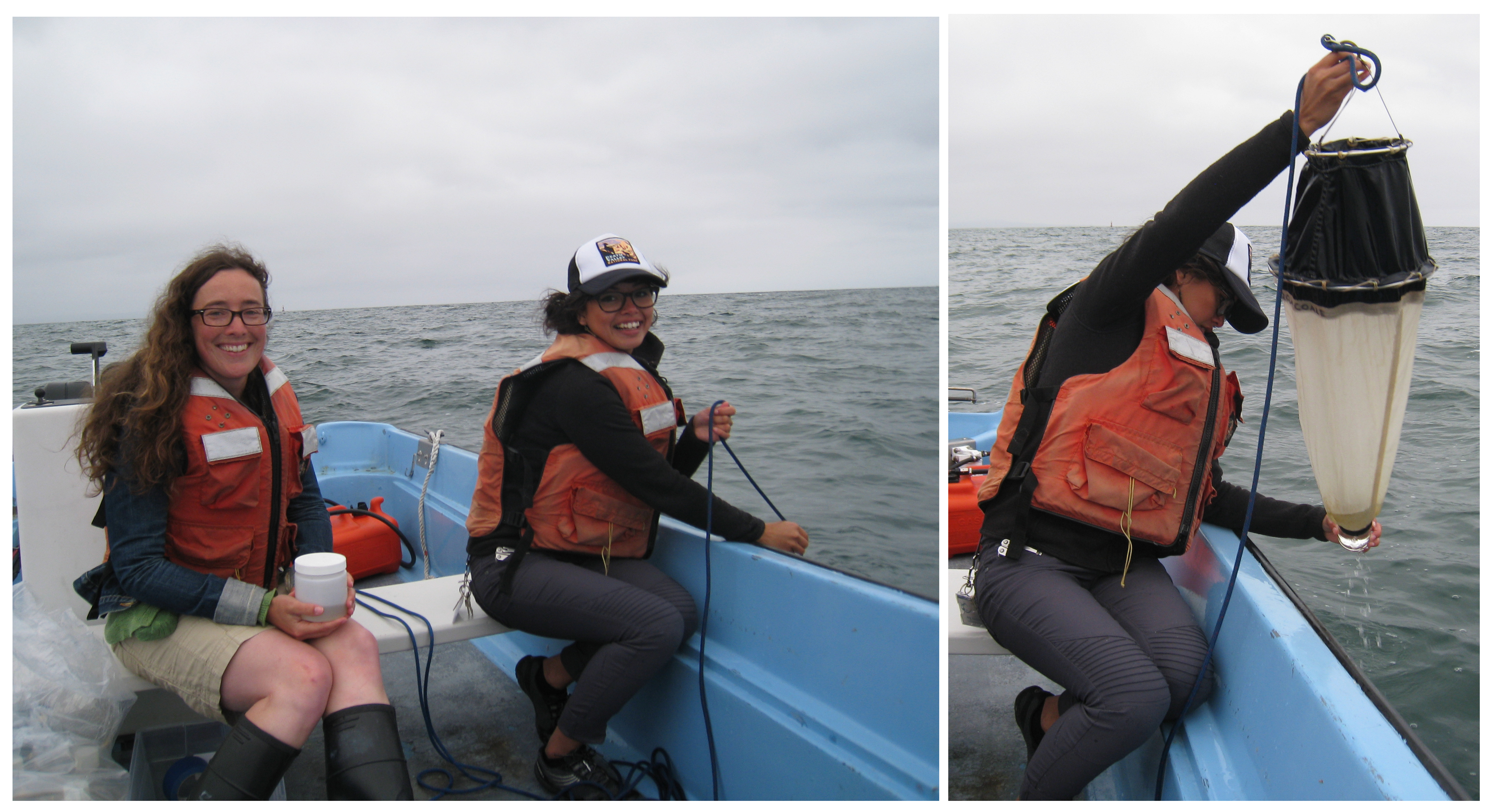
collecting seawater and a plankton net tow on an MLML boston whaler offshore of Moss Landing

Then I used a roller table to make aggregates.

In the lab, we used microscopy techniques to quantify the community composition of the aggregates and the corresponding surface water.

Aggregation_experiment_REU


The data was then used to identify if diatoms differentially aggregate and if morphology was driving differential aggregation. Throughout this process I was able to become familiar with over 20 diatom genera, using their shape and colony formation as identifiers.

(Note from Colleen: Melia estimates that she counted >21,000 individual diatom cells this summer!  She found significantly different phytoplankton compositions in aggregates compared to the total community in seawater, suggesting that some genera tend to be incorporated into aggregates more than others.  Melia plans to present this data at a conference later this year.)


Early summer particle flux at the New England continental shelf break

Last week I visited Melissa Omand’s lab at the University of Rhode Island to analyze sediment trap samples collected on the R/V Endeavor.  Unfortunately I was not able to go on the cruise, but I was still lucky enough to look at the exciting samples they brought back.

June_geltrap_jars

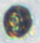
The types of samples collected on this cruise were very similar to those we collected in November.  Sediment traps collected sinking particles in the water column.  Above you can see how the number of sinking particles decreased with depth. These are images of the polyacrylamide gel jars placed in the bottom of traps at 4 depths spanning the upper mesopelagic zone.  The 60 meter trap was full of zooplankton fecal pellets (the long stringy particles).  At 150 meters, fluffy diatom aggregates appeared (mostly containing Pseudo-nitzschia), but sinking fecal pellets were also still abundant.  At high magnification, it became apparent that the traps were also chock-full of tiny little particle which turned out to be individual sinking coccolithophore cells. They are only about 10 micrometers big, and difficult to image clearly, but you can just make out the circular coccolith plates covering the outside of this cell .

Cocco

Although there is still much analysis to be done, these first observations are already exciting and represent a very different particle export environment than what we observed last November, which was dominated by large organic aggregates and large diatom cells.

On a related note, the URI Inner Space Center created several short videos about the research cruise last November.  Here are the films explaining the sediment traps.

Meg Estapa explaining the neutrally buoyant sediment traps:

Me, explaining the particle work:


Pat Kelly explaining the surface-tethered sediment trap array and in-situ pumps:

Today at the microscope (05/27/2016)

The main reason I enjoy working at the microscope is that I never know exactly what I will see.  Today while counting a preserved phytoplankton sample collected at the Rhode Island shelf break I spotted these cells:

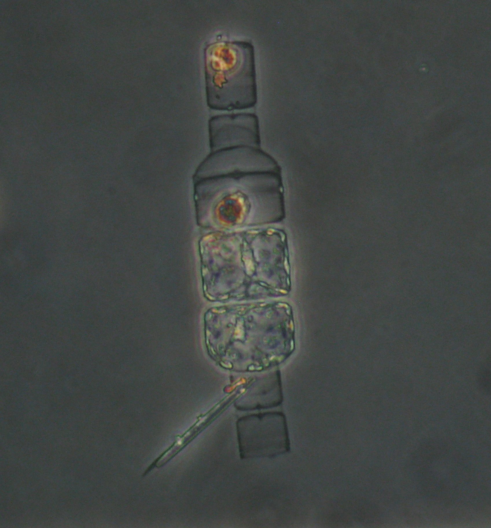
Detonula auxospores

This is a chain of diatom cells (genus: Detonula, I think) undergoing a life cycle stage that allows them to get bigger.  Diatom cells get smaller every time they divide.  At some point, the cells are too small to divide anymore and must form either sexual stages or auxospores to get big again.  In this photo the top cell in the chain is relatively small and does not appear to be vegetative (i.e. able to divide).  The second cell down the chain has a small top half and a much larger bottom half.  This cell formed an auxospore.  The third and forth cells down the chain are large and contain cytoplasm and chlorophyll (the yellow pigment).  These cells are probably the daughter (and grand-daughter) cells of that auxospore.  Because the cells are in a chain, it is possible to see the history of this life cycle response.

I found a very similar image of Detonula auxospores here.